US4829000A - Reconstituted basement membrane complex with biological activity - Google Patents
Reconstituted basement membrane complex with biological activity Download PDFInfo
- Publication number
- US4829000A US4829000A US06/867,027 US86702786A US4829000A US 4829000 A US4829000 A US 4829000A US 86702786 A US86702786 A US 86702786A US 4829000 A US4829000 A US 4829000A
- Authority
- US
- United States
- Prior art keywords
- extract
- buffer
- cells
- basement membrane
- collagen
- Prior art date
- Legal status (The legal status is an assumption and is not a legal conclusion. Google has not performed a legal analysis and makes no representation as to the accuracy of the status listed.)
- Expired - Lifetime
Links
Images


Classifications
-
- C—CHEMISTRY; METALLURGY
- C12—BIOCHEMISTRY; BEER; SPIRITS; WINE; VINEGAR; MICROBIOLOGY; ENZYMOLOGY; MUTATION OR GENETIC ENGINEERING
- C12N—MICROORGANISMS OR ENZYMES; COMPOSITIONS THEREOF; PROPAGATING, PRESERVING, OR MAINTAINING MICROORGANISMS; MUTATION OR GENETIC ENGINEERING; CULTURE MEDIA
- C12N5/00—Undifferentiated human, animal or plant cells, e.g. cell lines; Tissues; Cultivation or maintenance thereof; Culture media therefor
- C12N5/0018—Culture media for cell or tissue culture
-
- A—HUMAN NECESSITIES
- A61—MEDICAL OR VETERINARY SCIENCE; HYGIENE
- A61K—PREPARATIONS FOR MEDICAL, DENTAL OR TOILETRY PURPOSES
- A61K35/00—Medicinal preparations containing materials or reaction products thereof with undetermined constitution
- A61K35/12—Materials from mammals; Compositions comprising non-specified tissues or cells; Compositions comprising non-embryonic stem cells; Genetically modified cells
- A61K35/48—Reproductive organs
- A61K35/50—Placenta; Placental stem cells; Amniotic fluid; Amnion; Amniotic stem cells
-
- G—PHYSICS
- G01—MEASURING; TESTING
- G01N—INVESTIGATING OR ANALYSING MATERIALS BY DETERMINING THEIR CHEMICAL OR PHYSICAL PROPERTIES
- G01N33/00—Investigating or analysing materials by specific methods not covered by groups G01N1/00 - G01N31/00
- G01N33/48—Biological material, e.g. blood, urine; Haemocytometers
- G01N33/50—Chemical analysis of biological material, e.g. blood, urine; Testing involving biospecific ligand binding methods; Immunological testing
- G01N33/5005—Chemical analysis of biological material, e.g. blood, urine; Testing involving biospecific ligand binding methods; Immunological testing involving human or animal cells
- G01N33/5008—Chemical analysis of biological material, e.g. blood, urine; Testing involving biospecific ligand binding methods; Immunological testing involving human or animal cells for testing or evaluating the effect of chemical or biological compounds, e.g. drugs, cosmetics
- G01N33/5014—Chemical analysis of biological material, e.g. blood, urine; Testing involving biospecific ligand binding methods; Immunological testing involving human or animal cells for testing or evaluating the effect of chemical or biological compounds, e.g. drugs, cosmetics for testing toxicity
- G01N33/5017—Chemical analysis of biological material, e.g. blood, urine; Testing involving biospecific ligand binding methods; Immunological testing involving human or animal cells for testing or evaluating the effect of chemical or biological compounds, e.g. drugs, cosmetics for testing toxicity for testing neoplastic activity
-
- C—CHEMISTRY; METALLURGY
- C12—BIOCHEMISTRY; BEER; SPIRITS; WINE; VINEGAR; MICROBIOLOGY; ENZYMOLOGY; MUTATION OR GENETIC ENGINEERING
- C12N—MICROORGANISMS OR ENZYMES; COMPOSITIONS THEREOF; PROPAGATING, PRESERVING, OR MAINTAINING MICROORGANISMS; MUTATION OR GENETIC ENGINEERING; CULTURE MEDIA
- C12N2500/00—Specific components of cell culture medium
- C12N2500/70—Undefined extracts
- C12N2500/80—Undefined extracts from animals
- C12N2500/84—Undefined extracts from animals from mammals
-
- Y—GENERAL TAGGING OF NEW TECHNOLOGICAL DEVELOPMENTS; GENERAL TAGGING OF CROSS-SECTIONAL TECHNOLOGIES SPANNING OVER SEVERAL SECTIONS OF THE IPC; TECHNICAL SUBJECTS COVERED BY FORMER USPC CROSS-REFERENCE ART COLLECTIONS [XRACs] AND DIGESTS
- Y10—TECHNICAL SUBJECTS COVERED BY FORMER USPC
- Y10S—TECHNICAL SUBJECTS COVERED BY FORMER USPC CROSS-REFERENCE ART COLLECTIONS [XRACs] AND DIGESTS
- Y10S435/00—Chemistry: molecular biology and microbiology
- Y10S435/8215—Microorganisms
- Y10S435/948—Microorganisms using viruses or cell lines
Definitions
- the present invention relates generally to basement membrane complex. More particularly, the present invention relates to reconstituted, basement-membrane-derived extracellular substratum (matrigel) which polymerizes on heating and promotes cell growth and differentiation in vitro and in vivo.
- basement-membrane-derived extracellular substratum matrigel
- Basement membranes are thin, but continuous sheets that separate epithelium from stroma and surround nerves, muscle fibers, smooth muscle cells and fat cells.
- Basement membranes comprise type IV collagen, the glycoproteins laminin, entactin, nidogen and heparan sulfate proteoglycans. In various studies, these materials show a codistribution within both the lamina densa and its extensions across the lamina lucida. In the electron microscope, the components appear as a network of 5 nm wide cords and their codistribution suggests that the formation of basement membrane occurs through the interaction of various components. Type IV collagen molecules form intermolecular disulfide bonds and associate in a continuous network which can be visualized in basement membranes digested with plasmin (Inoue et al., J. Cell Biol. 97, 1524-1537, 1983).
- an object of the present invention to provide a reconstituted, basement-membrane-derived extracellular composition (matrigel) capable of polymerizing on heating and forming a three dimensional matrix which promotes cell growth and differentiation in vitro and in vivo.
- matrigel basement-membrane-derived extracellular composition
- An other object of the present invention is to provide a method of determining metastatic potential of tumor cells and of isolating metastatic tumor cells.
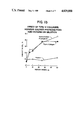
- FIG. 1 shows the effect of type IV collagen, heparan sulfate proteoglycan, and heparin on the gelation of basement membrane components from the basement membrane extract. Increasing amounts of each component were added to 100 ⁇ l of the extract and incubated for one hour at 35° in 0.15M NaCl, 0.05M Tris-HCl, pH 7.4. The samples were then centrifuged and insoluble material was dissolved in sample buffer. Equal aliquots of the samples were electrophoresed in 5% acrylamide. Densitometric scans of negatives of photographs of the gels were used to quantitate the amount of protein pelleted.
- A The effect of type IV collagen on the amount of total protein in the gel.
- FIG. 2 shows the effect of time and added type IV collagen on the gelation of the basement membrane extract. The conditions are similar to those described in the legend for FIG. 1. This figure compares gelation in the presence ( ⁇ ) and absence (O) of type IV collagen (50 ⁇ g);
- FIG. 3 shows the effect of temperature on the gelation of extracts of basement membrane.
- the experiment was carried out in the presence of type IV collagen (50 ⁇ g) as described in the legend for FIG. 2. Gelation was stopped by centrifugation at the times indicated;
- FIG. 4 shows the ability of the basement membrane extract to re-gel following dissolution of the gel.
- the first lane designated “whole extract” demonstrates the components in the starting material.
- the "first gel” designates the components in the gel formed in the presence of type IV collagen.
- the material present in the gel formed in the absence of type IV collagen (not shown) was solubilized for 20 minutes in 2.0M guanidine, dialyzed against 0.05M Tris-HCl, pH 7.4, containing 0.15M NaCl and allowed to regel in the absence (not shown) and presence of type IV collagen ("designated 2nd gel”). The cycle was repeated two additional times ("3rd” and "4th gels”). Shown are equal aliquots of the gels electrophoresed in a 5% acrylamide gel;
- FIG. 5 shows Sepharose 4B column chromatography of the 2.0M urea extract. Two ml of the whole extract equilibrated in either 2M urea, 0.15M NaCl, 0.05M Tris, pH 7.4 (dissociative), or 0.5M NaCl, 0.05M Tris, pH 7.4 (associative), were placed on a Sepharose 4B column (2 ⁇ 60 cm) equilibrated in the corresponding buffer (A). Aliquots of the designated fractions from the extract chromatographed in associative (B) or dissociative (C) conditions were analyzed by SDS polyacrylamide gels.
- FIG. 6 shows electron micrographs of reconstituted gels and an authentic basement membrane.
- A Gel formed in the absence of added type IV collagen or heparan sulfate proteoglycan. The gel consists of dispersed segments with occasional interconnections.
- B Gel formed in the presence of added type IV collagen and heparan sulfate proteoglycan. The edge of the gel is at the top. The gel consists of an interconnected network; the network is made up of structures which are similar in width to the lamina densa part of native basement membranes. These lamina densa-like structures vary somewhat in thickness.
- FIG. 7 shows the effect of the basement membrane gel on the morphology and differentiation of B16C3 melanoma cells in culture.
- A Morphology and assessment of melanogenesis by the cells.
- B Direct view of the dishes. The gel at the edge has been deflected to show that the cells are attached to it;
- FIG. 8 is Coomassie blue stain of 2.0M urea extract of placenta after heparin affinity chromatography showing the purification of laminin;
- FIG. 9 shows the effect of human placental matrigel on neuronal process formation. The results show that the human matrigel strongly promotes neurite outgrowth
- FIG. 10 is a diagramatic representation of Tumor Cell Invasiveness Assay using matrigel.
- FIG. 11 is a diagramatic representation of Invasive (Metastatic) Cell Selection using matrigel.
- a basement-membrane-derived composition comprising a biologically active polymerizable extract containing in parts by weight about 60-85% laminin, 5-30% collagen IV, 1-10% nidogen, 1-10% heparan sulfate proteoglycan and 1-5% entactin.
- biologically active as used herein means capable of supporting normal growth and differentiation of various cell types when cultured including epithelial cells.
- the reconstituted matrix promotes the growth and differentation of a variety of cells.
- the reconstituted basement membrane gel of the present invention is an excellent substrate for epithelial cells in culture.
- the matrigel of the present invention has also been demonstrated to promote cell adhesion, growth and differentiation of a multiplicity of cells including neurons, hepatocytes, sertoli cells, hair follicles, thyroid cells and the like.
- sertoli cells cultured within the gel have been subsequently transplanted back into the animal with good survival and maturation of the spermatids.
- the composition of the present invention has also been found to promote nerve regeneration (optic and sciatic) in vivo and allows for organ reconstitution as well. Preparing matrigel using an extract from human placenta also reduces the possibility of immunological interaction or rejection when such matrigel is used in humans.
- the basement membrane matrix was extracted with 0.5M NaCl in 0.05M Tris-HCl, pH 7.4.
- Laminin was isolated from the 0.5M NaCl extract as described by Timpl et al, supra.
- the residue of tumor tissue from lathyritic animals was extracted with 2.0M guanidine in 0.05M Tris-HCl, pH 7.4, followed by an extraction with the same buffer containing 0.005M dithiothreitol to solubilize the type IV collagen (Kleinman et al, supra).
- Low density heparan sulfate proteoglycan was purified from 6.0M urea extracts of the tumor by ion exchange chromatography followed by cesium chloride density centrifugation and molecular sieve column chromatography (Hassell et al, supra). Heparin was obtained from Sigma Chemical Company.
- Unfractionated extracts of the basement membrane matrix were prepared by treating the tissue which had been washed with high salt with an equal volume (1 ml/gm) of 2M urea, 0.05 M Tris-HCl, pH 7.4, overnight at 4° and centrifuging at 10,000g for 30 minutes. The residue was washed once with the same volume of buffer. Then the extract and wash were combined, dialyzed against 0.15M NaCl in 0.05M Tris-HCl, pH 7.4 (TBS), and centrifuged to remove a small amount of insoluble material. The supernatant fraction was stored at -20° C. in small aliquots and used in the reconstitution assays described below.
- this extract was found to contain laminin (3.5 mg/ml), type IV collagen (0.1 mg/ml) and heparan sulfate proteoglycan (0.1 mg/ml). Entactin, nidogen, and other minor components were also present.
- the extract was dialyzed into 0.5M NaCl, 0.05M Tris-HCl, pH 7.4, and centrifuged to remove insoluble material.
- Reconstitution Assays - Gelation was carried out in a centrifuge tube to which 0.05-0.1 ml of the 2M urea extract was added in physiological buffer. Purified components dissolved in 0.15M NaCl, 0.05 Tris-HCl, pH 7.4, were added to the extract or were incubated together at the concentrations indicated. The final volume was made up to 0.5 or 1.0 ml with 0.15M NaCl, 0.05M Tris-HCl, pH 7.4, and the samples were incubated for 1 hour at 35°C.
- Insoluble material was isolated by centrifugation and the pellets were dissolved in sample buffer and electrophoresed in either 5% or 7.5% acrylamide under reducing conditions (Laemmli, 1970, Nature - London, 227:680-682). Each experiment was repeated a minimum of three times. The total amount of protein in the precipitate was determined by the standard Lowry procedure. The amount of nidogen and entactin in the gel was related to the total amount of material present in the 400K band of laminin by scanning negatives of photographs of the gels in a Helena densitometer (Quick Scan Model, Helena Lab Corp., Beaumont, Tex.).
- Entactin and nidogen were identified based on their migration in SDS gels and cross reactivity in Western blot analyses with suitable antibodies.
- Type IV collagen in the gel was quantitated using 14 C-labeled type IV collagen and heparan sulfate proteoglycan was quantitated using 35 S-sulfate labeled material of known specific activities in separate but parallel experiments.
- Rotary Shadowing The 2.0M urea extract equilibrated in 0.5M NaCl, 0.05M Tris-HCl, pH 7.4, was placed on a Sepharose 4B column. An aliquot (30 ⁇ l ) of the peak fraction (0.1 mg/ml) eluting from the column was diluted with 300 ⁇ l of 0.155M ammonium acetate, pH 7.4, and 600 ⁇ l of glycerol. For rotary shadowing, the mixture was sprayed onto mica, shadowed with platinum-palladium, carbon coated, and examined in a JEOL 100C electron microscope.
- Ultrastructure of Reconstituted Components - The gel was prepared essentially as described above. Briefly, 0.2 ml of the extract was incubated alone or in the presence of type IV collagen and heparan sulfate proteoglycan overnight at 35° C. The gel was isolated by centrifugation and then fixed in 2.5% glutaraldehyde, treated with 1% OsO 4 , block stained with 2% uranyl acetate, and dehydrated. The gel was then processed through Epon (Ladd Research Industries, Inc., Burlington, Vt.; LX-112 resin) for electron microscopy. Thin sections were stained with uranyl acetate and lead citrate, and examined in a JEOL 100° C. electron microscope. Thin sections of rat kidney tubule basement membranes were obtained as described by Laurie et al, (Am. J. Anat. 169:463-481; 1984).
- Cell Culture - B16C3 cells were cultured either directly on tissue culture plastic or on a 1 mm thick basement membrane gel in a mixture of F12 medium and DMEM (Dulbecco's modified Eagle's medium, lacking phenol red for visualization of the pigmentation of the cells) containing glutamine, antibiotics, 20 mM tyrosine and 5% fetal calf serum. After one week, the cells were photographed.
- DMEM Dulbecco's modified Eagle's medium, lacking phenol red for visualization of the pigmentation of the cells
- the assembly of basement membrane components was analyzed using purified basement membrane components as well as unfractionated extracts of basement membrane.
- Purified type IV collagen, laminin and heparan sulfate proteoglycan formed a flocculent precipitate when incubated under physiological conditions for one hour at 35°.
- a gel formed when urea extracts of basement membrane are dialyzed against physiological saline and then warmed to 35° for one hour.
- the components of the gel were isolated by centrifugation and examined by SDS gel electrophoresis. As shown in FIG.
- the amount of laminin, entactin, and nidogen present in the gel increased in proportion to the amount of type IV collagen added until 50-60% of the material in the extract was incorporated into the gel.
- Heparan sulfate proteoglycan also caused increasing amounts of basement membrane components to precipitate (FIG. 1B). Separation by gel electrophoresis and quantitation of the major components in the gel indicated that constant ratios of laminin, entactin, and nidogen are obtained in the presence of added type IV collagen (FIG. 1A)or of heparan sulfate proteoglycan (FIG. 1B).
- type IV collagen 3 H-labeled type IV collagen of known specific activity was used and the amount of 3H-label in the precipitate was used as a measure of type IV collagen.
- the heparan sulfate proteoglycan cannot be visualized in the gels and 35 S-labeled heparan sulfate proteoglycan was used.
- the stability of the gel to dissolutioh was examined by using various solvents.
- the gel was not dissolved by cold aqueous salt but was partially dissolved by acidic solutions (Table 1) and completely dissolved in guanidine or in urea solutions. This suggests that the components are linked by relatively strong non-covalent bonds.
- Table 1 acidic solutions
- guanidine-dissolved gel was dialyzed against physiological buffers and warmed in the presence of type IV collagen, gel-like structures were reconstituted. This process could be repeated several times with similar proportions of laminin, nidogen, and entactin being deposited at each step as determined by SDS polyacrylamide gels (FIG. 4). In the presence of added type IV collagen, reformation of the gel occurred more rapidly and greater amounts of the components were deposited.
- the ultrastructure of the reconstituted basement membrane either with or without type IV collagen and heparan sulfate proteoglycan was also examined.
- the gel consisted of numerous widely separated thin, filamentous aggregates (FIG. 6A).
- the addition of type IV collagen or of heparan sulfate proteoglycan plus type IV collagen (FIG. 6B) resulted in the formation of thin sheets which were interconnected (FIG. 6B) or were confluent.
- the individual segments of the network had an average width similar to that of the lamina densa of kidney tubule basement membrane (FIG. 6C).
- the matrigel (reconstituted basement membrane) was used to coat the surfaces of bacteriological petri dishes and tested as a substrate for the growth and differentiation of a variety of cells at different laboratories.
- Melanoma cells (B16C3) showed considerable differences in morphology when grown on the basement membrane gel as compared to tissue culture surfaces (FIG. 7). Further, there was a much earlier and more extensive pigmentation of the cells on this substrate. Studies of other cells showed that endothelial cells formed tube-like structures on the gel and that hepatocytes survived longer on basement membrane gel substrates than on tissue culture plates or on type I collagen. In vivo, the basement membrane gel was found to promote peripheral nerve regeneration (Madison et al, 1985. Exptl. Neurol., 88:767-772).
- the reconstituted basement membrane is a biologically active substrate which induces diverse cellular responses. Since it can support cell adhesion, growth and differentiation beyond that known for the individual components, without being bound to any theory it is postulated that the reconstituted basement membrane gel contains these molecules in a unique and active conformation.
- the procedure is based on about 100 g of tumor and all steps are carried out at 4° C. unless indicated otherwise.
- Last dialysis steps should be against media salts such as DMEM or Dulbecco-Vogt or the like.
- Collagen IV can be optionally added at this stage to the liquid phase in an amount ranging from about 0.1 to 1 mg/ml depending on the desired consistency or strength of the polymerized gel matrix. The thicker gels have been found to be more durable.
- the gel as a cell culture substratum, add about 3 ml of suitable growth medium on top of the polymerized gel obtained from step 18 and inoculate the medium with the dispersions of the cells which are desired to be grown.
- suitable growth medium will depend on the type of the cell which is desired to be grown; specific standard growth medium and conditions (e.g. Co 2 concentration, temperature, pH and the like) for different types of cells being well known in the art.
- An alternate procedure for promoting the growth of some cell types is to inoculate or disperse the cells in the cold liquid extract just before polymerization in step 18 and then proceed with polymerization and subsequent steps the same as described in step 19.
- hair follicle, sertoli cells and the like are apt to be better cultured if first dispersed in the liquid phase prior to polymerization whereas epithelial cells, exocrine acinar cells, sciatic nerve cell, spinal cord neuron, thyroid organ culture, and the like are better cultured on top of the polymerized gel.
- Extracts comparable in composition and in biological activity can also be obtained from human placenta using a process similar to that used for the EHS mouse tumor described herein. However, since placenta is not composed of pure basement membrane like the EHS mouse tumor. an additional step is necessary as described hereunder:
- Placenta is then washed and homogenized in about 3.4M NaCl in 0.05M Tris-HCl, pH 7.4 containing standard protease inhibitors such as phenylmethyl sulfonyl fluoride; n-ethylmaleimide EDTA, pepstatin and the like.
- tissue residue is extracted overnight at about 4° C. with an equal volume (g/ml) of 0.5M NaCl in 0.05M Tris-HCl, pH 7.4.
- tissue residue is extracted overnight at about 4° C. with an equal volume (g/ml) of 2.0M urea in 0.05M Tris-HCl, pH 7.4.
- Both the 0.5M NaCl extract and the 2.0M urea extract are dialyzed against 0.02M sodium phosphate buffer, pH 7.4 overnight at 4° C. and the dialyzed samples are separately chromatographed on a heparin Sepharose column equilibrated in 0.02M sodium phosphate buffer, pH 7.4, containing 0.15M NaCl. The bound material is eluted with 1.0M NaCl and dialyzed into Eagle's minimal essential medium.
- placental extract before and after heparin sepharose chromatography were compared for the purity of the extract.
- the 0.5M NaCl and 2.0M urea extracts and the bound materials from the heparin column were dialyzed against water, lyophillized, and electrophoresed in SDS polyacrylamide gels.
- the samples were then stained with Coomassie blue for a profile on the protein content and immunoreacted with anti-laminin antibodies after transfer to nitrocellulose.
- the biological activity of this material on neurite outgrowth was tested using NG108-15 neuroblastoma plus glioma hybrid cells in culture. These cells respond rapidly (within 2 hours) to the extracts as well as to the heparin bound material by sending out long neuritic processes (FIG. 9).
- the material to be tested is added in Eagle's minimal essential medium lacking serum or some other culture medium along with freshly dissociated cells. After two hours on tissue culture plastic, extended processes are observed in the cells exposed to the placental material. Thus, the placenta materials have comparable activity to the murine tumor material in stimulating neurite process development.
- tumor cells In order for all tumor cells to metastasize, they must enter the blood stream and then exit from it to grow at a distant site. Tumor cells must therefore adhere to, degrade, and migrate through endothelial basement membranes in order to metastasize. These steps are critical in tumor cell metastasis.
- a unique in vitro assay to measure these critical steps in the invasion process has now been devised. The assay is fast, quantitative, reproducible, and distinguishes between nonmetastatic and metastatic cells. This assay employs the murine reconstituted basement membrane described herein.
- a porous filter (Nucleopore) is placed inside a blind well Boyden chamber.
- the lower compartment contains an attractant such as fibroblast conditioned medium or laminin.
- Fifty microliters of murine basement membrane extract are placed on top of the filter in the upper compartment and allowed to polymerize at 37° C.
- cells in Eagle's minimal essential medium or some other suitable culture medium are added to the upper well and the entire chamber is incubated at 37° C. in 95% air, 5% CO 2 for 5 hours.
- the invasive cells adhere to the matrix, degrade the matrix, and migrate through the matrix and the porous holes in the filter. This process is diagrammatically shown in FIG. 10.
- the number of cells which have invaded the matrix can be quantitated on the lower side of the filter, for example by direct counting in a microscope after the cells have been stained with DifQuick (Harelco). Alternatively, if the cells are radiolabeled, they can be measured directly in a scintillation counter.
- Highly invasive tumor cells can also be selected for and obtained in pure form based on their ability to adhere, degrade, and migrate through the reconstituted basement membrane.
- the murine basement membrane extract is placed on a tissue culture dish (0.5 ml/35 mm diameter dish) and allowed to polymerize for 30 minutes at 37° C.
- the cells are plated in a sterile manner in complete culture medium as required for the growth of the specific cells. After two days, the invasive cells attach to, degrade, and migrate through the matrix to the surface of the plastic dish where they are concentrated. This is shown in FIG. 11.
- the invasive cells on the plastic surface can be recovered after removal of the reconstituted basement membrane gel.
- the selected cells are the parent lines.
- the selected cells are those from the parent line which have chemoinvaded the matrigel, been isolated and grown. Upon retesting their ability to invade the matrigel, they are much more invasive. Data are expressed as number of cell aggregates which have invaded the matrigel ⁇ 1 S.D.
Abstract
Description
TABLE 1 ______________________________________ SOLUBILIZATION OF RECONSTITUTED BASEMENT MEMBRANES Solvent % Solubilized ______________________________________ 0.15M NaCl 0 0.5M NaCl 0 0.5 M HAc 43 1.0 M urea +Dithiothreitol 40 2.0 M urea 73 2.0 M Guanidine 97 ______________________________________ All solutions except the 0.5 M HAc were buffered with 0.05 M TrisHCl at p 7.4. The solubilization was carried out at 24° for 20 minutes. After 20 minutes with frequent vortexing, the solutions were centrifuged and the pellets were redissolved and electrophoresed in SDS gels with a reducing agent. The relative amount of material in the pellets was determined by scanning the SDS gels.
              TABLE II
______________________________________
Tumorgenicity and Invasiveness of Human and
Murine Cell Lines
                  Tumor
                  Formation
                         Invasiveness
                  In Vivo
                         In Vitro
______________________________________
10T1/2 fibroblasts  No       No
NIH 3T3 fibroblasts No       No
NIH 3T3 transfected with RAS
                    Yes      Yes
NIH 3T3 transfected with MOS
                    Yes      Yes
NIH 3T3 transfected with SSV
                    Yes      Yes
NIH 3T3 transfected with MMSV
                    Yes      Yes
B16 F1 melanoma     Yes      Yes
B16 F10 melanoma    Yes      Yes
B16 BL6 melanoma    Yes      Yes
B16 Br2 melanoma    Yes      Yes
K-1735 melanoma Cl 10
                    No       No
K-1735 melanoma Cl 10
                    Yes      Yes
MC-180 epidermoid carcinoma
                    Yes      Yes
A-204 Rhabdomyosarcoma
                    Yes      Yes
PA-1 Teratocarcinoma
                    Yes      Yes
PC-3 prostate carcinoma
                    Yes      Yes
MALME 3m Melamona   Yes      Yes
SW 620 Colon Adenocarcinoma
                    Yes      Yes
MCF-7 breast carcinoma
                    No       No
MCF-7 breast carcinoma &
                    Yes      Yes
estradiol
MCF-7 breast carcinoma &
                    Yes      Yes
ras
______________________________________
 Cells were assayed for 5 hours in the Boyden Chamber assay described
 above. Cell lines which had less than 5 cells/field on the lower surface
 of the filter were considered non invasive (No), whereas cell lines with
 10 cells or more/field on the lower surface were considered invasive
 (Yes). The ability of the cells lines to form tumors in vivo is available
 in published literature.
              TABLE III
______________________________________
Number of Cell Aggregates Invading Matrigel
Before and After Selection in Matrigel
Cells            # of aggregates
______________________________________
Unselected cells
Cl 10 (non metastatic)
                  2 ± 1
Cl 3 (low metastatic)
                  6 ± 1
M2 (high metastatic)
                 16 ± 1
Selected cells
Cl 10            19 ± 1
M2               19 ± 3
______________________________________
Claims (11)
Priority Applications (6)
| Application Number | Priority Date | Filing Date | Title |
|---|---|---|---|
| US06/867,027 US4829000A (en) | 1985-08-30 | 1986-05-27 | Reconstituted basement membrane complex with biological activity |
| AT86111635T ATE66247T1 (en) | 1985-08-30 | 1986-08-22 | RESTORED BASAL MEMBRANE COMPLEX WITH BIOLOGICAL ACTIVITY. |
| EP86111635A EP0218065B1 (en) | 1985-08-30 | 1986-08-22 | Reconstituted basement membrane complex with biological activity |
| DE8686111635T DE3680851D1 (en) | 1985-08-30 | 1986-08-22 | RESTORED BASAL MEMBRANE COMPLEX WITH BIOLOGICAL ACTIVITY. |
| CA000516643A CA1291432C (en) | 1985-08-30 | 1986-08-22 | Reconstituted basement membrane complex with biological activity |
| JP61203534A JPH0740932B2 (en) | 1985-08-30 | 1986-08-29 | Cell culture composition and use thereof |
Applications Claiming Priority (2)
| Application Number | Priority Date | Filing Date | Title |
|---|---|---|---|
| US77140985A | 1985-08-30 | 1985-08-30 | |
| US06/867,027 US4829000A (en) | 1985-08-30 | 1986-05-27 | Reconstituted basement membrane complex with biological activity |
Related Parent Applications (1)
| Application Number | Title | Priority Date | Filing Date |
|---|---|---|---|
| US77140985A Continuation-In-Part | 1985-08-30 | 1985-08-30 |
Related Child Applications (1)
| Application Number | Title | Priority Date | Filing Date |
|---|---|---|---|
| US07/291,623 Division US5158874A (en) | 1985-08-30 | 1988-12-29 | Determining metastic potential of tumor cells and isolating metastic tumor cells |
Publications (1)
| Publication Number | Publication Date |
|---|---|
| US4829000A true US4829000A (en) | 1989-05-09 |
Family
ID=27118460
Family Applications (1)
| Application Number | Title | Priority Date | Filing Date |
|---|---|---|---|
| US06/867,027 Expired - Lifetime US4829000A (en) | 1985-08-30 | 1986-05-27 | Reconstituted basement membrane complex with biological activity |
Country Status (6)
| Country | Link |
|---|---|
| US (1) | US4829000A (en) |
| EP (1) | EP0218065B1 (en) |
| JP (1) | JPH0740932B2 (en) |
| AT (1) | ATE66247T1 (en) |
| CA (1) | CA1291432C (en) |
| DE (1) | DE3680851D1 (en) |
Cited By (159)
| Publication number | Priority date | Publication date | Assignee | Title |
|---|---|---|---|---|
| WO1991006303A1 (en) * | 1989-10-27 | 1991-05-16 | Case Western Reserve University | Inhibition of cell growth by keratan sulfate, chondroitin sulfate, dermatan sulfate and other glycans |
| WO1991015245A1 (en) * | 1990-03-30 | 1991-10-17 | THE UNITED STATES OF AMERICA, as represented by THE SECRETARY, UNITED STATES DEPARTMENT OF COMMERCE | Method and composition for growing tumors from few cells |
| WO1992017498A1 (en) * | 1991-03-26 | 1992-10-15 | The State Of Oregon Acting By And Through The State Board Of Higher Education On Behalf Of The Oregon Health Sciences University | Product and method for improving keratinocyte adhesion to the dermis |
| US5175103A (en) * | 1991-10-21 | 1992-12-29 | Trustees Of University Of Pennsylvania | Preparation of pure cultures of post-mitotic human neurons |
| US5354666A (en) * | 1991-08-01 | 1994-10-11 | Thomas Jefferson University | Mammalian cell line expressing basement membrane proteins |
| WO1995020041A1 (en) * | 1994-01-21 | 1995-07-27 | The Regents Of The University Of California | Immortalized human myoepithelial cells and their uses |
| US5512657A (en) * | 1988-12-12 | 1996-04-30 | Bainbridge Sciences, Inc. | Detection of complexes which include basement membrane components as diagnostic of cancer and other diseases |
| US5541076A (en) * | 1988-12-12 | 1996-07-30 | Bard Diagnostic Sciences, Inc. | Methods for determining the invasiveness of a bladder tumor |
| US5547997A (en) * | 1991-10-01 | 1996-08-20 | Chemisches Laboratorium Dr. Kurt Richter Gmbh | Plant-derived cosmetic composition and method of treatment of skin |
| US5605938A (en) * | 1991-05-31 | 1997-02-25 | Gliatech, Inc. | Methods and compositions for inhibition of cell invasion and fibrosis using dextran sulfate |
| US5695998A (en) * | 1995-02-10 | 1997-12-09 | Purdue Research Foundation | Submucosa as a growth substrate for islet cells |
| US5705178A (en) * | 1991-05-31 | 1998-01-06 | Gliatech, Inc. | Methods and compositions based on inhibition of cell invasion and fibrosis by anionic polymers |
| US5759534A (en) * | 1994-04-13 | 1998-06-02 | Research Corporation Technologies, Inc. | Methods of treating disease using sertoli cells and allografts or xenografts |
| US5770562A (en) * | 1991-03-26 | 1998-06-23 | Oregon Health Sciences University | Products and methods for improving keratinocyte adhesion to the dermis |
| US5776747A (en) * | 1994-07-20 | 1998-07-07 | Cytotherapeutics, Inc. | Method for controlling the distribution of cells within a bioartificial organ using polycthylene oxide-poly (dimethylsiloxane) copolymer |
| US5834029A (en) * | 1994-07-20 | 1998-11-10 | Cytotherapeutics, Inc. | Nerve guidance channel containing bioartificial three-dimensional hydrogel extracellular matrix derivatized with cell adhesive peptide fragment |
| US5843431A (en) * | 1994-07-20 | 1998-12-01 | Cytotherapeutics, Inc. | Controlling proliferation of cells before and after encapsulation in a bioartificial organ by gene transformation |
| US5869266A (en) * | 1990-03-06 | 1999-02-09 | The United States Of America As Represented By The Department Of Health And Human Services | Human olfactory neuron cultures to diagnose Alzheimer's disease |
| WO1999032607A1 (en) * | 1997-12-23 | 1999-07-01 | Purdue Research Foundation | Biomaterial derived from follicle basement membranes |
| US6074874A (en) * | 1997-08-29 | 2000-06-13 | University Of Pittsburgh | Epithelial cell cultures for in vitro testing |
| US6133236A (en) * | 1991-03-26 | 2000-10-17 | Oregon Health Sciences University | Products and methods for improving keratinocyte adhesion to the dermis |
| US6264992B1 (en) * | 1998-02-27 | 2001-07-24 | Purdue Research Foundation | Submucosa as a growth substrate for cells |
| US6326019B1 (en) | 1997-02-28 | 2001-12-04 | Scheffer C. G. Tseng | Grafts made from amniotic membrane; methods of separating, preserving, and using such grafts in surgeries |
| WO2002020825A1 (en) | 2000-09-09 | 2002-03-14 | The Research Foundation Of State University Of New York | Method and compositions for isolating metastatic cancer cells, and use in measuring metastatic potential of a cancer thereof |
| US6375989B1 (en) | 1996-12-10 | 2002-04-23 | Purdue Research Foundation | Submucosa extracts |
| US6379710B1 (en) * | 1996-12-10 | 2002-04-30 | Purdue Research Foundation | Biomaterial derived from vertebrate liver tissue |
| US20020123141A1 (en) * | 2000-12-06 | 2002-09-05 | Hariri Robert J. | Method of collecting placental stem cells |
| US6479072B1 (en) | 1999-02-11 | 2002-11-12 | The General Hospital Corporation | Microfabricated membranes and matrices |
| US6482410B1 (en) * | 1994-09-16 | 2002-11-19 | The Scripps Research Institute | Cytotactin derivatives that stimulate attachment and neurite outgrowth, and methods of making same |
| US6485723B1 (en) | 1995-02-10 | 2002-11-26 | Purdue Research Foundation | Enhanced submucosal tissue graft constructs |
| US6495364B2 (en) * | 1995-05-23 | 2002-12-17 | Neurotech, S.A. | Mx-1 conditionally immortalized cells |
| US20020197718A1 (en) * | 2001-06-06 | 2002-12-26 | Becton, Dickinson And Company | Method of providing a substrate with a ready-to-use, uniformly distributed extracellular matrix |
| US20030014126A1 (en) * | 2001-06-28 | 2003-01-16 | Patel Umesh H. | Graft prosthesis devices containing renal capsule collagen |
| US20030032179A1 (en) * | 2000-12-06 | 2003-02-13 | Hariri Robert J. | Post-partum mammalian placenta, its use and placental stem cells therefrom |
| US6649160B1 (en) | 1995-03-13 | 2003-11-18 | University Of South Florida | Sertoli cells as transplantation facilitator for cell transplantation |
| US20030216812A1 (en) * | 2002-05-02 | 2003-11-20 | Badylak Stephen F. | Vascularization enhanced graft constructs |
| US20030216811A1 (en) * | 2002-05-02 | 2003-11-20 | Badylak Stephen F. | Vascularization enhanced graft constructs |
| US20030215426A1 (en) * | 2002-04-02 | 2003-11-20 | William Marsh Rice University | Redifferentiated cells for repairing cartilage defects |
| US6667176B1 (en) * | 2000-01-11 | 2003-12-23 | Geron Corporation | cDNA libraries reflecting gene expression during growth and differentiation of human pluripotent stem cells |
| JP2004500093A (en) * | 2000-01-27 | 2004-01-08 | メディジェーン アクチェンゲゼルシャフト | Apparatus and method for measuring contraction of three-dimensional matrix body and cell tissue |
| US20040006395A1 (en) * | 2002-05-02 | 2004-01-08 | Badylak Stephen F. | Vascularization enhanced graft constructs |
| US6696270B2 (en) | 1996-12-10 | 2004-02-24 | Purdue Research Foundation | Gastric submucosal tissue as a novel diagnostic tool |
| US6706520B2 (en) | 2001-06-13 | 2004-03-16 | Kehan Han | Assessment of invasive potential of tumor cells |
| US6740501B2 (en) * | 2000-09-27 | 2004-05-25 | Becton, Dickinson And Company | Coated membrane for assessing the invasive capacity of a cell |
| US20040176855A1 (en) * | 2003-03-07 | 2004-09-09 | Acell, Inc. | Decellularized liver for repair of tissue and treatment of organ deficiency |
| US20040175366A1 (en) * | 2003-03-07 | 2004-09-09 | Acell, Inc. | Scaffold for cell growth and differentiation |
| US20040187877A1 (en) * | 2002-12-04 | 2004-09-30 | Badylak Stephen F. | Method for repair of liver tissue |
| US20040191226A1 (en) * | 2002-12-04 | 2004-09-30 | Badylak Stephen F. | Method for repair of body wall |
| US6815203B1 (en) * | 1999-06-23 | 2004-11-09 | Joslin Diabetes Center, Inc. | Methods of making pancreatic islet cells |
| US20050019419A1 (en) * | 2002-01-11 | 2005-01-27 | Badylak Stephen Francis | Biomaterial derived from vertebrate liver tissue |
| US20050025838A1 (en) * | 2003-06-25 | 2005-02-03 | Badylak Stephen F. | Conditioned compositions for tissue restoration |
| JP2005065696A (en) * | 2003-08-20 | 2005-03-17 | Becton Dickinson & Co | Modified reconstituted basement membrane composition for assay system |
| US20050089516A1 (en) * | 2002-06-18 | 2005-04-28 | Shiseido Company, Ltd. | Skin vitalizing composition for external use anti-aging preparation |
| WO2005042048A2 (en) | 2003-10-22 | 2005-05-12 | Encelle, Inc. | Bioactive hydrogel compositions for regenerating connective tissue |
| WO2004076631A3 (en) * | 2003-02-21 | 2005-05-12 | Uab Research Foundation | Biologically active native biomatrix composition |
| US20050175705A1 (en) * | 2003-08-20 | 2005-08-11 | Becton, Dickinson And Company | Modified reconstituted basement membrane composition for assay system |
| US20050208627A1 (en) * | 2003-09-18 | 2005-09-22 | Bowdish Katherine S | Elicitation of antibodies to self peptides in mice by immunization with dendritic cells |
| US20050244843A1 (en) * | 2001-11-16 | 2005-11-03 | Wen-Tien Chen | Blood test prototypes and methods for the detection of circulating tumor and endothelial cells |
| US20050276792A1 (en) * | 2004-03-26 | 2005-12-15 | Kaminski Joseph K | Systems and methods for providing a stem cell bank |
| WO2005121316A1 (en) * | 2004-06-11 | 2005-12-22 | Bernard O'brien Institute Of Microsurgery | Tissue material and muscle derived matrix |
| US20060121008A1 (en) * | 2002-06-18 | 2006-06-08 | Eisai Co., Ltd. | Primarily cultured adipocytes for gene therapy |
| WO2005043121A3 (en) * | 2003-10-31 | 2006-08-24 | Vitatex Inc | Blood test prototypes and methods for the detection of circulating tumor and endothelial cells |
| US20060251630A1 (en) * | 2002-12-24 | 2006-11-09 | An-Go-Gen Inc. | Encapsulated cell therapy |
| US20060252149A1 (en) * | 2005-05-06 | 2006-11-09 | Orion Biosolutions, Inc. | Cns cells in vitro |
| US20060263417A1 (en) * | 2005-05-10 | 2006-11-23 | Lelkes Peter I | Electrospun blends of natural and synthetic polymer fibers as tissue engineering scaffolds |
| US20070054378A1 (en) * | 2003-09-18 | 2007-03-08 | Alexion Pharmaceuticals, Inc. | Elicitation of antibodies to self peptides in mice by immunization with dendritic cells |
| US20070077265A1 (en) * | 2003-11-07 | 2007-04-05 | Klueh Ulrike W | Article tissue systems and uses thereof |
| US20070190165A1 (en) * | 2005-10-21 | 2007-08-16 | Brey Eric M | Tissue-specific basement membrane gels |
| US7259140B2 (en) | 2003-03-28 | 2007-08-21 | Thomas Jefferson University | Heparin-binding peptides and uses thereof |
| US20080032401A1 (en) * | 2005-12-29 | 2008-02-07 | James Edinger | Placental stem cell populations |
| US20080145348A1 (en) * | 1998-11-06 | 2008-06-19 | Sertoli Technologies, Inc. | Production of a biological factor and creation of an immunologically privileged environment using genetically altered sertoli cells |
| US20080206343A1 (en) * | 2007-02-12 | 2008-08-28 | Edinger James W | Hepatocytes and Chondrocytes from Adherent Placental StemCells; And CD34+ ,CD45- Placental Stem Cell-Enriched Cell Populations |
| US20080213389A1 (en) * | 2005-06-17 | 2008-09-04 | Drexel University | Three-Dimensional Scaffolds for Tissue Engineering Made by Processing Complex Extracts of Natural Extracellular Matrices |
| US20080274184A1 (en) * | 2004-03-31 | 2008-11-06 | Hunt James B | Ecm-Based Graft Material |
| US20080289395A1 (en) * | 2007-05-23 | 2008-11-27 | Universal Scientific Industrial Co., Ltd. | Testing machine |
| US20080299582A1 (en) * | 1998-10-23 | 2008-12-04 | Geron Corporation | Culture System for Rapid Expansion of Human Embryonic Stem Cells |
| US20090136553A1 (en) * | 2007-09-25 | 2009-05-28 | Gerlach Jorg C | Triggerably dissolvable hollow fibers for controlled delivery |
| US20090162331A1 (en) * | 2003-07-03 | 2009-06-25 | Sertoli Technologies, Inc. | Compositions containing sertoli cells and myoid cells and use thereof in cellular transplants |
| US20090215434A1 (en) * | 2003-09-11 | 2009-08-27 | Cvon Innovations Limited | Method and system for distributing data to mobile devices |
| US20090264678A1 (en) * | 2007-08-27 | 2009-10-22 | John Robert Lockemeyer | Carrier, a process for preparing the carrier, an olefin epoxidation catalyst, a process for preparing the catalyst, and a process for the production of an olefin oxide, a 1,2-diol, a 1,2-diol ether, or an alkanolamine |
| US20100047351A1 (en) * | 2008-08-20 | 2010-02-25 | Andy Zeitlin | Treatment of stroke using isolated placental cells |
| US7682803B2 (en) | 2005-10-13 | 2010-03-23 | Anthrogenesis Corporation | Immunomodulation using placental stem cells |
| US7700090B2 (en) | 2002-02-13 | 2010-04-20 | Anthrogenesis Corporation | Co-culture of placental stem cells and stem cells from a second source |
| US20100228335A1 (en) * | 2009-03-04 | 2010-09-09 | John Schorgl | Stents modified with material comprising amnion tissue and corresponding processes |
| US20100310529A1 (en) * | 2007-12-03 | 2010-12-09 | Irina Aizman | Extracellular matrix from pluripotent cells |
| US20110003387A1 (en) * | 2009-07-02 | 2011-01-06 | Abbot Stewart | Method of producing erythrocytes without feeder cells |
| US20110020418A1 (en) * | 2009-07-22 | 2011-01-27 | Bosley Jr Rodney W | Particulate Tissue Graft with Components of Differing Density and Methods of Making and Using the Same |
| US20110020420A1 (en) * | 2009-07-22 | 2011-01-27 | Bosley Jr Rodney W | Variable Density Tissue Graft Composition and Methods of Making and Using the Same |
| US20110076329A1 (en) * | 1996-08-23 | 2011-03-31 | Cook William A | Graft prosthesis, material and methods |
| US20110172159A1 (en) * | 2008-09-05 | 2011-07-14 | Coelho De Sampaio Tatiana Lobo | Proteic acid polymer, production processes, use of proteic acid polymer, pharmaceutical composition and method of treatment |
| WO2011099007A1 (en) | 2010-02-10 | 2011-08-18 | Nayacure Therapeutics Ltd. | Pharmaceutical compositions and methods for the treatment and prevention of cancer |
| WO2012027592A2 (en) * | 2010-08-25 | 2012-03-01 | Lifenet Health | Basement membrane compositions and applications thereof |
| US8263065B2 (en) | 2007-09-28 | 2012-09-11 | Anthrogenesis Corporation | Tumor suppression using human placental perfusate and human placenta-derived intermediate natural killer cells |
| WO2013003234A1 (en) * | 2011-06-28 | 2013-01-03 | Trevigen, Inc. | Tumor extract |
| US8367409B2 (en) | 2008-11-19 | 2013-02-05 | Anthrogenesis Corporation | Amnion derived adherent cells |
| US8377484B1 (en) | 2004-05-06 | 2013-02-19 | Maria V. Tsiper | Tumor encapsulation for prevention and treatment of metastatic cancer disease |
| EP2594635A1 (en) | 2011-11-18 | 2013-05-22 | Univercell Biosolutions | Method for generating primate cardiovascular progenitor cells for clinical and drug cells testing use from primate embryonic stem cells or embryonic-like state cells, and their applications |
| US8460650B2 (en) | 2007-02-12 | 2013-06-11 | Anthrogenesis Corporation | Treatment of inflammatory diseases using placental stem cells |
| WO2013096650A2 (en) | 2011-12-23 | 2013-06-27 | Pioneer Surgical Technology | Continuous matrix with osteoconductive particles dispersed therein, method of forming thereof, and method of regenerating bone therewith |
| US8562973B2 (en) | 2010-04-08 | 2013-10-22 | Anthrogenesis Corporation | Treatment of sarcoidosis using placental stem cells |
| WO2013162828A1 (en) | 2012-04-27 | 2013-10-31 | The United States Of America, As Represented By The Secretary, Department Of Health & Human Services | Use of cpg oligonucleotides co-formulated with an antibiotic to accelarate wound healing |
| US8617535B2 (en) | 2002-11-26 | 2013-12-31 | Anthrogenesis Corporation | Cytotherapeutics, cytotherapeutic units and methods for treatments using them |
| US20140072510A1 (en) * | 2012-09-13 | 2014-03-13 | Northwestern University | Synthetic Scaffolds for Metastasis Detection |
| US8728805B2 (en) | 2008-08-22 | 2014-05-20 | Anthrogenesis Corporation | Methods and compositions for treatment of bone defects with placental cell populations |
| EP2743345A1 (en) | 2012-12-13 | 2014-06-18 | IMBA-Institut für Molekulare Biotechnologie GmbH | Three dimensional heterogeneously differentiated tissue culture |
| EP2774935A1 (en) | 2009-10-30 | 2014-09-10 | NTF Therapeutics, Inc. | Improved neurturin molecules |
| US20140294780A1 (en) * | 2013-04-02 | 2014-10-02 | University Of Florida Research Foundation, Inc. | Compositions and methods for induction and modulation of angiogenesis and methods and assays for identifying angiogenesis modulators |
| US8926964B2 (en) | 2010-07-13 | 2015-01-06 | Anthrogenesis Corporation | Methods of generating natural killer cells |
| US8969315B2 (en) | 2010-12-31 | 2015-03-03 | Anthrogenesis Corporation | Enhancement of placental stem cell potency using modulatory RNA molecules |
| US9040035B2 (en) | 2011-06-01 | 2015-05-26 | Anthrogenesis Corporation | Treatment of pain using placental stem cells |
| US9121007B2 (en) | 2010-01-26 | 2015-09-01 | Anthrogenesis Corporatin | Treatment of bone-related cancers using placental stem cells |
| US9161955B2 (en) | 2005-09-27 | 2015-10-20 | Tissuetech, Inc. | Amniotic membrane preparations and purified compositions and therapy for scar reversal and inhibition |
| US9161954B2 (en) | 2005-09-27 | 2015-10-20 | Tissuetech, Inc. | Amniotic membrane preparations and purified compositions and anti-angiogenesis treatment |
| US9200253B1 (en) | 2007-08-06 | 2015-12-01 | Anthrogenesis Corporation | Method of producing erythrocytes |
| US9254302B2 (en) | 2010-04-07 | 2016-02-09 | Anthrogenesis Corporation | Angiogenesis using placental stem cells |
| US9327010B2 (en) | 2005-04-25 | 2016-05-03 | Massachusetts Institute Of Technology | Compositions and methods for promoting hemostasis and other physiological activities |
| US9415084B2 (en) | 2007-03-14 | 2016-08-16 | Arch Biosurgery, Inc. | Treatment of leaky or damaged tight junctions and enhancing extracellular matrix |
| WO2016146893A1 (en) | 2015-03-17 | 2016-09-22 | University Of Oulu | Human tumor based extracellular matrix for cell studies in vitro |
| EP3095470A1 (en) | 2008-02-07 | 2016-11-23 | Shahar Cohen | Compartmental extract compositions for tissue engineering |
| US9511113B2 (en) | 2006-04-25 | 2016-12-06 | Massachusetts Institute Of Technology | Compositions and methods for affecting movement of contaminants, bodily fluids or other entities, and/or affecting other physiological conditions |
| WO2017007325A1 (en) | 2015-07-09 | 2017-01-12 | Mimetas B.V. | Barrier function measurements |
| US9682044B2 (en) | 2011-06-10 | 2017-06-20 | Tissuetech, Inc. | Methods of processing fetal support tissues, fetal support tissue powder products, and uses thereof |
| EP3190176A1 (en) | 2016-01-11 | 2017-07-12 | IMBA-Institut für Molekulare Biotechnologie GmbH | Method for tissue culture development on scaffold and differentiated tissue culture |
| WO2017132627A2 (en) | 2016-01-29 | 2017-08-03 | Achaogen, Inc. | Screening methods for identifying antibodies that bind cell surface epitopes |
| WO2017155399A1 (en) | 2016-03-09 | 2017-09-14 | Mimetas B.V. | Double tubular structures |
| US9763983B2 (en) | 2013-02-05 | 2017-09-19 | Anthrogenesis Corporation | Natural killer cells from placenta |
| US9789157B2 (en) | 2005-04-25 | 2017-10-17 | Arch Biosurgery, Inc. | Compositions for prevention of adhesions and other barrier applications |
| WO2017184601A1 (en) | 2016-04-19 | 2017-10-26 | The United States Of America, As Represented By The Secretary, Department Of Health And Human Services | Use of gram negative species to treat atopic dermatitis |
| US9821022B2 (en) | 2013-08-22 | 2017-11-21 | Arch Biosurgery, Inc. | Implantable meshes for controlling the movement of fluids |
| WO2017216113A2 (en) | 2016-06-15 | 2017-12-21 | Mimetas B.V. | Cell culture device and methods |
| WO2018023129A1 (en) * | 2016-07-29 | 2018-02-01 | Zpredicta, Inc. | Screening of immuno-modulatory therapies |
| US9925221B2 (en) | 2011-09-09 | 2018-03-27 | Celularity, Inc. | Treatment of amyotrophic lateral sclerosis using placental stem cells |
| EP3338549A1 (en) | 2007-11-20 | 2018-06-27 | Pioneer Surgical Orthobiologics, Inc. | Cryopreservation of cells using cross-linked bioactive hydrogel matrix particles |
| WO2018140827A1 (en) | 2017-01-27 | 2018-08-02 | Achaogen, Inc. | Reporter microorganisms and uses thereof |
| US10104880B2 (en) | 2008-08-20 | 2018-10-23 | Celularity, Inc. | Cell composition and methods of making the same |
| EP3395942A1 (en) | 2017-04-25 | 2018-10-31 | IMBA-Institut für Molekulare Biotechnologie GmbH | Bi- or multi-differentiated organoid |
| US10130288B2 (en) | 2013-03-14 | 2018-11-20 | Cell and Molecular Tissue Engineering, LLC | Coated sensors, and corresponding systems and methods |
| WO2018229251A1 (en) | 2017-06-16 | 2018-12-20 | Imba - Institut Für Molekulare Biotechnologie Gmbh | Blood vessel organoid, methods of producing and using said organoids |
| WO2019048689A1 (en) | 2017-09-11 | 2019-03-14 | Imba - Institut Für Molekulare Biotechnologie Gmbh | Tumor organoid model |
| WO2019050015A1 (en) | 2017-09-08 | 2019-03-14 | 国立研究開発法人理化学研究所 | Cell aggregate including retinal tissue and production method therefor |
| WO2019083904A1 (en) | 2017-10-23 | 2019-05-02 | Chan Zuckerberg Biohub, Inc. | Measurement of afucosylated igg fc glycans and related treatment methods |
| US10293005B2 (en) | 2016-04-19 | 2019-05-21 | The United States Of America, As Represented By The Secretary, Department Of Health And Human Services | Use of gram negative species to treat atopic dermatitis |
| US10377986B2 (en) * | 2014-02-12 | 2019-08-13 | The Board Of Regents Of The University Of Texas System | Three-dimensional silk fibroin scaffold culture retaining functional salivary cells and promoting salivary tissue-specific ECM synthesis |
| US10405961B2 (en) | 2013-03-14 | 2019-09-10 | Cell and Molecular Tissue Engineering, LLC | Coated surgical mesh, and corresponding systems and methods |
| WO2020109421A1 (en) | 2018-11-28 | 2020-06-04 | Mimetas B.V. | Device for assessing mechanical strain induced in or by cells |
| WO2020152272A1 (en) | 2019-01-23 | 2020-07-30 | United Kingdom Research And Innovation | Choroid plexus organoids and methods for production thereof |
| EP3689971A1 (en) | 2019-02-04 | 2020-08-05 | Real Research Sp. z o.o. | Protein hydrogel, preparation method and use thereof |
| WO2020169551A1 (en) | 2019-02-19 | 2020-08-27 | Miltenyi Biotec B.V. & Co. KG | Cell culture medium and method for generation of epithelial organoids from epithelial stem cells |
| US10973841B2 (en) | 2018-05-11 | 2021-04-13 | Forte Subsidiary, Inc. | Compositions for the treatment of skin conditions |
| WO2021137222A1 (en) | 2019-12-31 | 2021-07-08 | Ichilov Tech Ltd. | Methods of treating atopic dermatitis |
| US11124756B2 (en) | 2014-01-22 | 2021-09-21 | Zpredicta, Inc. | Method and apparatus for isolating invasive and metastatic cells for evaluating therapeutics and prediction of metastatic capacity |
| WO2022008733A1 (en) | 2020-07-09 | 2022-01-13 | Mimetas B.V. | Microfluidic cell culture device |
| US11478574B2 (en) | 2013-10-16 | 2022-10-25 | Purdue Research Foundation | Collagen compositions and methods of use |
| CN115261302A (en) * | 2022-07-20 | 2022-11-01 | 创芯国际生物科技(广州)有限公司 | Matrigel and preparation method and application thereof |
| US11590265B2 (en) | 2015-02-23 | 2023-02-28 | Biotissue Holdings Inc. | Apparatuses and methods for treating ophthalmic diseases and disorders |
| US11707492B2 (en) | 2016-01-29 | 2023-07-25 | Biotissue Holdings Inc. | Fetal support tissue products and methods of use |
| US11739291B2 (en) | 2017-04-25 | 2023-08-29 | Purdue Research Foundation | 3-dimensional (3D) tissue-engineered muscle for tissue restoration |
| EP4286513A1 (en) | 2022-06-03 | 2023-12-06 | IMBA-Institut für Molekulare Biotechnologie GmbH | Triple tissue culture fusion |
| US11919941B2 (en) | 2015-04-21 | 2024-03-05 | Purdue Research Foundation | Cell-collagen-silica composites and methods of making and using the same |
Families Citing this family (6)
| Publication number | Priority date | Publication date | Assignee | Title |
|---|---|---|---|---|
| US5037656A (en) * | 1986-12-04 | 1991-08-06 | Millipore Corporation | Porous membrane having hydrophilic and cell growth promotions surface and process |
| US5731417A (en) * | 1994-04-25 | 1998-03-24 | Becton, Dickinson And Company | Cell culture substrates and method of making |
| DE10010113B4 (en) * | 2000-03-03 | 2009-05-07 | Wolfgang Dr. Schatton | Native sponge collagen, process for its isolation and its use, as well as native nanoparticulate sponge collagen, process for its preparation and its use |
| DE102007026639A1 (en) | 2007-06-06 | 2008-12-11 | Stefan-Andreas Ulrich | Producing animal leather skin substitutes, comprises carrying out animal in-vitro growing of epithelial cells; laminating cultivated partly active cells on sterilized substrate; and cultivating the cells in translucent incubator system |
| CA2948348A1 (en) * | 2014-05-08 | 2015-11-12 | The University Of Florida Research Foundation, Inc. | Sustained release angiogenesis modulating compositions and methods for induction and modulation of angiogenesis |
| US20230138007A1 (en) * | 2020-04-08 | 2023-05-04 | Ichimaru Pharcos Co., Ltd. | Unit for angiogenesis promotion and/or nerve regeneration |
Citations (2)
| Publication number | Priority date | Publication date | Assignee | Title |
|---|---|---|---|---|
| US4280954A (en) * | 1975-07-15 | 1981-07-28 | Massachusetts Institute Of Technology | Crosslinked collagen-mucopolysaccharide composite materials |
| US4642292A (en) * | 1979-10-29 | 1987-02-10 | Albert Einstein College Of Medicine Of Yeshiva University, A Division Of Yeshiva University | Method for isolation of connective tissue biomatrix |
-
1986
- 1986-05-27 US US06/867,027 patent/US4829000A/en not_active Expired - Lifetime
- 1986-08-22 DE DE8686111635T patent/DE3680851D1/en not_active Expired - Lifetime
- 1986-08-22 AT AT86111635T patent/ATE66247T1/en not_active IP Right Cessation
- 1986-08-22 CA CA000516643A patent/CA1291432C/en not_active Expired - Lifetime
- 1986-08-22 EP EP86111635A patent/EP0218065B1/en not_active Expired - Lifetime
- 1986-08-29 JP JP61203534A patent/JPH0740932B2/en not_active Expired - Lifetime
Patent Citations (2)
| Publication number | Priority date | Publication date | Assignee | Title |
|---|---|---|---|---|
| US4280954A (en) * | 1975-07-15 | 1981-07-28 | Massachusetts Institute Of Technology | Crosslinked collagen-mucopolysaccharide composite materials |
| US4642292A (en) * | 1979-10-29 | 1987-02-10 | Albert Einstein College Of Medicine Of Yeshiva University, A Division Of Yeshiva University | Method for isolation of connective tissue biomatrix |
Non-Patent Citations (24)
| Title |
|---|
| Evercooren et al., (1982), Journ. of Neuroscience Res., 8:179 193. * |
| Evercooren et al., (1982), Journ. of Neuroscience Res., 8:179-193. |
| Grant, D. S. et al., Chem. Abstr., 104:49361y. * |
| Hassell et al., (1985), Develop. Mech.: Normal and Abnormal, pp. 75 86. * |
| Hassell et al., (1985), Develop. Mech.: Normal and Abnormal, pp. 75-86. |
| Hogan, B. L. M. et al., Chem. Abstr., 102:76352m. * |
| Kleinman et al., (1982), Biochem., 21(24):6188. * |
| Kleinman et al., (1983), Biochem., 22(21), pp. 4969 4974. * |
| Kleinman et al., (1983), Biochem., 22(21), pp. 4969-4974. |
| Martin et al., Chem. Abstr., 102:93811c. * |
| Martin, G. R. et al., (1984), Ciba Foundation Symposium, 108, 197 212. * |
| Martin, G. R. et al., (1984), Ciba Foundation Symposium, 108, 197-212. |
| McGarvey et al., (1984), Develop. Biol., 105:18 28. * |
| McGarvey et al., (1984), Develop. Biol., 105:18-28. |
| Reid et al., Methods Enzymol, 58:263 278, 1979. * |
| Reid et al., Methods Enzymol, 58:263-278, 1979. |
| Terranova et al., (1983), Proc. Natl. Acad. Sci. U.S.A., 80:444 448. * |
| Terranova et al., (1983), Proc. Natl. Acad. Sci. U.S.A., 80:444-448. |
| Terranova, V. P. et al., Science, 226:982 985, 1984. * |
| Terranova, V. P. et al., Science, 226:982-985, 1984. |
| Timpl et al., Eur. J. Biochem., 137:455 465, 1983. * |
| Timpl et al., Eur. J. Biochem., 137:455-465, 1983. |
| Woodley et al., (1983), Journ. Invest. Derm., 81:149 153. * |
| Woodley et al., (1983), Journ. Invest. Derm., 81:149-153. |
Cited By (348)
| Publication number | Priority date | Publication date | Assignee | Title |
|---|---|---|---|---|
| US5591830A (en) * | 1988-12-12 | 1997-01-07 | Bainbridge Sciences, Inc. | Detection of complexes which include basement membrane components as diagnostic of cancer and other diseases |
| US5591595A (en) * | 1988-12-12 | 1997-01-07 | Bainbridge Sciences, Inc. | Detection of complexes which include basement membrane components as diagnostic of cancer and other diseases |
| US5512657A (en) * | 1988-12-12 | 1996-04-30 | Bainbridge Sciences, Inc. | Detection of complexes which include basement membrane components as diagnostic of cancer and other diseases |
| US5541076A (en) * | 1988-12-12 | 1996-07-30 | Bard Diagnostic Sciences, Inc. | Methods for determining the invasiveness of a bladder tumor |
| WO1991006303A1 (en) * | 1989-10-27 | 1991-05-16 | Case Western Reserve University | Inhibition of cell growth by keratan sulfate, chondroitin sulfate, dermatan sulfate and other glycans |
| US5869266A (en) * | 1990-03-06 | 1999-02-09 | The United States Of America As Represented By The Department Of Health And Human Services | Human olfactory neuron cultures to diagnose Alzheimer's disease |
| WO1991015245A1 (en) * | 1990-03-30 | 1991-10-17 | THE UNITED STATES OF AMERICA, as represented by THE SECRETARY, UNITED STATES DEPARTMENT OF COMMERCE | Method and composition for growing tumors from few cells |
| WO1992017498A1 (en) * | 1991-03-26 | 1992-10-15 | The State Of Oregon Acting By And Through The State Board Of Higher Education On Behalf Of The Oregon Health Sciences University | Product and method for improving keratinocyte adhesion to the dermis |
| US5770562A (en) * | 1991-03-26 | 1998-06-23 | Oregon Health Sciences University | Products and methods for improving keratinocyte adhesion to the dermis |
| US6133236A (en) * | 1991-03-26 | 2000-10-17 | Oregon Health Sciences University | Products and methods for improving keratinocyte adhesion to the dermis |
| US5352668A (en) * | 1991-03-26 | 1994-10-04 | The State Of Oregon Acting By And Through The State Board Of Higher Education On Behalf Of Oregon Health Sciences University | Product and method for improving keratinocyte adhesion to the dermis |
| US5705178A (en) * | 1991-05-31 | 1998-01-06 | Gliatech, Inc. | Methods and compositions based on inhibition of cell invasion and fibrosis by anionic polymers |
| US6127348A (en) * | 1991-05-31 | 2000-10-03 | Gliatech, Inc. | Methods and compositions based on inhibition of cell invasion and fibrosis by anionic polymers |
| US5994325A (en) * | 1991-05-31 | 1999-11-30 | Gliatech Inc. | Methods and compositions based on inhibition of cell invasion and fibrosis by anionic polymers |
| US5605938A (en) * | 1991-05-31 | 1997-02-25 | Gliatech, Inc. | Methods and compositions for inhibition of cell invasion and fibrosis using dextran sulfate |
| US6020326A (en) * | 1991-05-31 | 2000-02-01 | Gliatech Inc. | Method for inhibition of bone growth by anionic polymers |
| US5705177A (en) * | 1991-05-31 | 1998-01-06 | Gliatech Inc. | Methods and compositions based on inhibition of cell invasion and fibrosis by anionic polymers |
| US6083930A (en) * | 1991-05-31 | 2000-07-04 | Gliatech Inc. | Methods and compositions based on inhibition of cell invasion and fibrosis by anionic polymers |
| US5354666A (en) * | 1991-08-01 | 1994-10-11 | Thomas Jefferson University | Mammalian cell line expressing basement membrane proteins |
| US5547997A (en) * | 1991-10-01 | 1996-08-20 | Chemisches Laboratorium Dr. Kurt Richter Gmbh | Plant-derived cosmetic composition and method of treatment of skin |
| US5654189A (en) * | 1991-10-21 | 1997-08-05 | Trustees Of The University Of Pennsylvania | Preparation of pure cultures of post-mitotic human neurons |
| US5175103A (en) * | 1991-10-21 | 1992-12-29 | Trustees Of University Of Pennsylvania | Preparation of pure cultures of post-mitotic human neurons |
| WO1993008266A1 (en) * | 1991-10-21 | 1993-04-29 | The Trustees Of The University Of Pennsylvania | Preparation of pure cultures of post-mitotic human neurons |
| WO1995020041A1 (en) * | 1994-01-21 | 1995-07-27 | The Regents Of The University Of California | Immortalized human myoepithelial cells and their uses |
| US5508188A (en) * | 1994-01-21 | 1996-04-16 | The Regents Of The University Of California | Method of growing cells in a mammal |
| US5759534A (en) * | 1994-04-13 | 1998-06-02 | Research Corporation Technologies, Inc. | Methods of treating disease using sertoli cells and allografts or xenografts |
| AU715177B2 (en) * | 1994-04-13 | 2000-01-20 | Research Corporation Technologies, Inc. | Methods of treating disease using sertoli cells and allografts or xenografts |
| US6156572A (en) * | 1994-07-20 | 2000-12-05 | Neurotech S.A. | Bioartificial extracellular matrix containing hydrogel matrix derivatized with cell adhesive peptide fragment |
| US5776747A (en) * | 1994-07-20 | 1998-07-07 | Cytotherapeutics, Inc. | Method for controlling the distribution of cells within a bioartificial organ using polycthylene oxide-poly (dimethylsiloxane) copolymer |
| US5858747A (en) * | 1994-07-20 | 1999-01-12 | Cytotherapeutics, Inc. | Control of cell growth in a bioartificial organ with extracellular matrix coated microcarriers |
| US5853717A (en) * | 1994-07-20 | 1998-12-29 | Cytotherapeutics, Inc. | Methods and compositions of growth control for cells encapsulated within bioartificial organs |
| US5843431A (en) * | 1994-07-20 | 1998-12-01 | Cytotherapeutics, Inc. | Controlling proliferation of cells before and after encapsulation in a bioartificial organ by gene transformation |
| US6392118B1 (en) | 1994-07-20 | 2002-05-21 | Neurotech S.A. | Mx-1 conditionally immortalized cells |
| US5935849A (en) * | 1994-07-20 | 1999-08-10 | Cytotherapeutics, Inc. | Methods and compositions of growth control for cells encapsulated within bioartificial organs |
| US5840576A (en) * | 1994-07-20 | 1998-11-24 | Cytotherapeutics, Inc. | Methods and compositions of growth control for cells encapsulated within bioartificial organs |
| US5834029A (en) * | 1994-07-20 | 1998-11-10 | Cytotherapeutics, Inc. | Nerve guidance channel containing bioartificial three-dimensional hydrogel extracellular matrix derivatized with cell adhesive peptide fragment |
| US5833979A (en) * | 1994-07-20 | 1998-11-10 | Cytotherapeutics, Inc. | Methods and compositions of growth control for cells encapsulated within bioartificial organs |
| US5795790A (en) * | 1994-07-20 | 1998-08-18 | Cytotherapeutics, Inc. | Method for controlling proliferation and differentiation of cells encapsulated within bioartificial organs |
| US6482410B1 (en) * | 1994-09-16 | 2002-11-19 | The Scripps Research Institute | Cytotactin derivatives that stimulate attachment and neurite outgrowth, and methods of making same |
| US7175841B2 (en) | 1995-02-10 | 2007-02-13 | Purdue Research Foundation | Enhanced submucosal tissue graft constructs |
| US5753267A (en) * | 1995-02-10 | 1998-05-19 | Purdue Research Foundation | Method for enhancing functional properties of submucosal tissue graft constructs |
| US5695998A (en) * | 1995-02-10 | 1997-12-09 | Purdue Research Foundation | Submucosa as a growth substrate for islet cells |
| US6087157A (en) * | 1995-02-10 | 2000-07-11 | Clarian Health Partners | Device and method for analyzing tumor cell invasion of an extracellular matrix |
| US20110165126A1 (en) * | 1995-02-10 | 2011-07-07 | Purdue Research Foundation | Enhanced Submucosal Tissue Graft Constructs |
| US20070141037A1 (en) * | 1995-02-10 | 2007-06-21 | Badylak Stephen F | Enhanced submucosal tissue graft constructs |
| US20030064056A1 (en) * | 1995-02-10 | 2003-04-03 | Badylak Stephen F. | Enhanced submucosal tissue graft constructs |
| US7771717B2 (en) * | 1995-02-10 | 2010-08-10 | Purdue Research Foundation | Enhanced submucosal tissue graft constructs |
| US6485723B1 (en) | 1995-02-10 | 2002-11-26 | Purdue Research Foundation | Enhanced submucosal tissue graft constructs |
| US5866414A (en) * | 1995-02-10 | 1999-02-02 | Badylak; Stephen F. | Submucosa gel as a growth substrate for cells |
| US6649160B1 (en) | 1995-03-13 | 2003-11-18 | University Of South Florida | Sertoli cells as transplantation facilitator for cell transplantation |
| US6495364B2 (en) * | 1995-05-23 | 2002-12-17 | Neurotech, S.A. | Mx-1 conditionally immortalized cells |
| US20110076329A1 (en) * | 1996-08-23 | 2011-03-31 | Cook William A | Graft prosthesis, material and methods |
| US8920516B2 (en) | 1996-08-23 | 2014-12-30 | Cook Biotech Incorporated | Graft prosthesis, material and methods |
| US20090123511A1 (en) * | 1996-12-10 | 2009-05-14 | Purdue Research Foundation | Biomaterial derived from vertebrate liver tissue |
| US6793939B2 (en) | 1996-12-10 | 2004-09-21 | Purdue Research Foundation | Biomaterial derived from vertebrate liver tissue |
| US8003131B2 (en) | 1996-12-10 | 2011-08-23 | Purdue Research Foundation | Biomaterial derived from vertebrate liver tissue |
| US7482025B2 (en) | 1996-12-10 | 2009-01-27 | Purdue Research Foundation | Biomaterial derived from vertebrate liver tissue |
| US6696270B2 (en) | 1996-12-10 | 2004-02-24 | Purdue Research Foundation | Gastric submucosal tissue as a novel diagnostic tool |
| US20040157323A1 (en) * | 1996-12-10 | 2004-08-12 | Badylak Stephen F. | Biomaterial derived from vertebrate liver tissue |
| US6375989B1 (en) | 1996-12-10 | 2002-04-23 | Purdue Research Foundation | Submucosa extracts |
| US20040157283A1 (en) * | 1996-12-10 | 2004-08-12 | Badylak Stephen F. | Gastric submucosal tissue as a novel diagnostic tool |
| US6379710B1 (en) * | 1996-12-10 | 2002-04-30 | Purdue Research Foundation | Biomaterial derived from vertebrate liver tissue |
| US8647677B2 (en) | 1996-12-10 | 2014-02-11 | Purdue Research Foundation | Gastric submucosal tissue as a novel diagnostic tool |
| US6326019B1 (en) | 1997-02-28 | 2001-12-04 | Scheffer C. G. Tseng | Grafts made from amniotic membrane; methods of separating, preserving, and using such grafts in surgeries |
| US6383805B1 (en) | 1997-08-29 | 2002-05-07 | University Of Pittsburgh | Epithelial cell cultures for in vitro testing |
| US6074874A (en) * | 1997-08-29 | 2000-06-13 | University Of Pittsburgh | Epithelial cell cultures for in vitro testing |
| US6485969B1 (en) | 1997-12-23 | 2002-11-26 | Purdue Research Foundation | Biomaterial derived from follicle basement membranes |
| WO1999032607A1 (en) * | 1997-12-23 | 1999-07-01 | Purdue Research Foundation | Biomaterial derived from follicle basement membranes |
| US20030012822A1 (en) * | 1998-02-27 | 2003-01-16 | Voytik-Harbin Sherry L. | Submucosa gel compositions |
| US20030012823A1 (en) * | 1998-02-27 | 2003-01-16 | Voytik-Harbin Sherry L. | Submucosa gel compositions |
| US6444229B2 (en) | 1998-02-27 | 2002-09-03 | Purdue Research Foundation | Submucosa gel compositions |
| US6264992B1 (en) * | 1998-02-27 | 2001-07-24 | Purdue Research Foundation | Submucosa as a growth substrate for cells |
| US7147871B2 (en) | 1998-02-27 | 2006-12-12 | Purdue Research Foundation | Submucosa gel compositions |
| US20080299582A1 (en) * | 1998-10-23 | 2008-12-04 | Geron Corporation | Culture System for Rapid Expansion of Human Embryonic Stem Cells |
| US8637311B2 (en) | 1998-10-23 | 2014-01-28 | Asterias Biotherapeutics, Inc. | Human embryonic stem cells genetically modified to contain a nucleic acid and cultured with fibroblast growth factor |
| US20100317101A1 (en) * | 1998-10-23 | 2010-12-16 | Geron Corporation | Culture System for Rapid Expansion of Human Embryonic Stem Cells |
| US10059939B2 (en) | 1998-10-23 | 2018-08-28 | Asterias Biotherapeutics, Inc. | Screening methods for human embryonic stem cells |
| US20100203633A1 (en) * | 1998-10-23 | 2010-08-12 | Ramkumar Mandalam | Culture System for Rapid Expansion of Human Embryonic Stem Cells |
| US8097458B2 (en) | 1998-10-23 | 2012-01-17 | Geron Corporation | Micro-carrier culture system for rapid expansion of human embryonic stem cells |
| US8951800B2 (en) | 1998-10-23 | 2015-02-10 | Asterias Biotherapeutics, Inc. | Primate pluripotent stem cell expansion without feeder cells and in the presence of FGF and matrigel or Engelbreth-Holm-Swarm tumor cell preparation |
| US20080145348A1 (en) * | 1998-11-06 | 2008-06-19 | Sertoli Technologies, Inc. | Production of a biological factor and creation of an immunologically privileged environment using genetically altered sertoli cells |
| US20030044395A1 (en) * | 1999-02-11 | 2003-03-06 | The General Hospital Corporation, A Massachusetts Corporation | Microfabricated membranes and matrices |
| US6479072B1 (en) | 1999-02-11 | 2002-11-12 | The General Hospital Corporation | Microfabricated membranes and matrices |
| US20050069529A1 (en) * | 1999-06-23 | 2005-03-31 | Joslin Diabetes Center, Inc., A Massachusetts Corporation | Methods of making pancreatic islet cells |
| US6815203B1 (en) * | 1999-06-23 | 2004-11-09 | Joslin Diabetes Center, Inc. | Methods of making pancreatic islet cells |
| US6667176B1 (en) * | 2000-01-11 | 2003-12-23 | Geron Corporation | cDNA libraries reflecting gene expression during growth and differentiation of human pluripotent stem cells |
| JP2004500093A (en) * | 2000-01-27 | 2004-01-08 | メディジェーン アクチェンゲゼルシャフト | Apparatus and method for measuring contraction of three-dimensional matrix body and cell tissue |
| EP1315829A1 (en) * | 2000-09-09 | 2003-06-04 | The Research Foundation of State University of New York | Method and compositions for isolating metastatic cancer cells, and use in measuring metastatic potential of a cancer thereof |
| EP1315829A4 (en) * | 2000-09-09 | 2005-02-23 | Res Foundation Ofstate Univers | Method and compositions for isolating metastatic cancer cells, and use in measuring metastatic potential of a cancer thereof |
| WO2002020825A1 (en) | 2000-09-09 | 2002-03-14 | The Research Foundation Of State University Of New York | Method and compositions for isolating metastatic cancer cells, and use in measuring metastatic potential of a cancer thereof |
| US7687241B2 (en) | 2000-09-09 | 2010-03-30 | The Research Foundation Of State University Of New York | Methods and compositions for isolating metastatic cancer cells, and use in measuring metastatic potential of a cancer thereof |
| US20100173402A1 (en) * | 2000-09-09 | 2010-07-08 | The Research Foundation Of State University Of New York | Methods and Compositions for Isolating Metastatic Cancer Cells, and Use in Measuring Metastatic Potential of a Cancer Thereof |
| US20030206901A1 (en) * | 2000-09-09 | 2003-11-06 | Wen-Tien Chen | Method and compositions for isolating metastatic cancer cells, and use in measuring metastatic potentatial of a cancer thereof |
| AU2011201368B2 (en) * | 2000-09-09 | 2013-08-15 | The Research Foundation Of State University Of New York | Method and compositions for isolating metastatic cancer cells, and use in measuring metastatic potential of a cancer thereof |
| US20050153342A1 (en) * | 2000-09-09 | 2005-07-14 | The Research Foundation Of State University Of New York | Methods and compositions for isolating metastatic cancer cells, and use in measuring metastatic potential of a cancer thereof |
| US7785810B2 (en) | 2000-09-09 | 2010-08-31 | The Research Foundation Of State University Of New York | Method and compositions for isolating metastatic cancer cells, and use in measuring metastatic potential of a cancer thereof |
| US8288116B2 (en) | 2000-09-09 | 2012-10-16 | The Research Foundation Of State University Of New York | Methods and compositions for isolating metastatic cancer cells, and use in measuring metastatic potential of a cancer thereof |
| US6740501B2 (en) * | 2000-09-27 | 2004-05-25 | Becton, Dickinson And Company | Coated membrane for assessing the invasive capacity of a cell |
| US8986984B2 (en) | 2000-12-06 | 2015-03-24 | Anthrogenesis Corporation | Method of propagating cells |
| US20100120015A1 (en) * | 2000-12-06 | 2010-05-13 | Hariri Robert J | Method of collecting placental stem cells |
| US20110217272A1 (en) * | 2000-12-06 | 2011-09-08 | Anthrogenesis Corporation | Treatment of radiation injury using placental stem cells |
| US7045148B2 (en) * | 2000-12-06 | 2006-05-16 | Anthrogenesis Corporation | Method of collecting placental stem cells |
| US20110217271A1 (en) * | 2000-12-06 | 2011-09-08 | Anthrogenesis Corporation | Treatment of diseases or disorders using placental stem cells |
| US20020123141A1 (en) * | 2000-12-06 | 2002-09-05 | Hariri Robert J. | Method of collecting placental stem cells |
| US20090053805A1 (en) * | 2000-12-06 | 2009-02-26 | Anthrogenesis Corporation | Post-partum mammalian placenta, its use and placental stem cells therefrom |
| US7976836B2 (en) | 2000-12-06 | 2011-07-12 | Anthrogenesis Corporation | Treatment of stroke using placental stem cells |
| US20090142831A1 (en) * | 2000-12-06 | 2009-06-04 | Anthrogenesis Corporation | Placental stem cells |
| US8057788B2 (en) | 2000-12-06 | 2011-11-15 | Anthrogenesis Corporation | Placental stem cell populations |
| US20040048372A1 (en) * | 2000-12-06 | 2004-03-11 | Anthrogenesis Corporation | Post-partum mammalian placenta, its use and placental stem cells therefrom |
| US9149569B2 (en) | 2000-12-06 | 2015-10-06 | Anthrogenesis Corporation | Treatment of diseases or disorders using placental stem cells |
| US8580563B2 (en) | 2000-12-06 | 2013-11-12 | Anthrogenesis Corporation | Placental stem cells |
| US7255879B2 (en) | 2000-12-06 | 2007-08-14 | Anthrogenesis Corporation | Post-partum mammalian placenta, its use and placental stem cells therefrom |
| US9387283B2 (en) | 2000-12-06 | 2016-07-12 | Anthrogenesis Corporation | Method of collecting placental stem cells |
| US8293223B2 (en) | 2000-12-06 | 2012-10-23 | Anthrogenesis Corporation | Treatment of organ injuries and burns using placental stem cells |
| US8545833B2 (en) | 2000-12-06 | 2013-10-01 | Anthrogenesis Corporation | Treatment of radiation injury using placental stem cells |
| US7468276B2 (en) | 2000-12-06 | 2008-12-23 | Anthrogenesis Corporation | Placental stem cells |
| US20030032179A1 (en) * | 2000-12-06 | 2003-02-13 | Hariri Robert J. | Post-partum mammalian placenta, its use and placental stem cells therefrom |
| US7541187B2 (en) | 2001-06-06 | 2009-06-02 | Becton, Dickinson And Company | Method of providing a substrate with a ready-to-use, uniformly distributed extracellular matrix |
| US20020197718A1 (en) * | 2001-06-06 | 2002-12-26 | Becton, Dickinson And Company | Method of providing a substrate with a ready-to-use, uniformly distributed extracellular matrix |
| US6706520B2 (en) | 2001-06-13 | 2004-03-16 | Kehan Han | Assessment of invasive potential of tumor cells |
| US7745217B2 (en) | 2001-06-28 | 2010-06-29 | Cook Biotech Incorporated | Graft prosthesis devices containing renal capsule collagen |
| US20070037283A1 (en) * | 2001-06-28 | 2007-02-15 | Cook Biotech Incorporated | Graft prosthesis devices containing renal capsule collagen |
| US7087089B2 (en) | 2001-06-28 | 2006-08-08 | Cook Biotech Incorporated | Graft prosthesis devices containing renal capsule collagen |
| US20030014126A1 (en) * | 2001-06-28 | 2003-01-16 | Patel Umesh H. | Graft prosthesis devices containing renal capsule collagen |
| US20050244843A1 (en) * | 2001-11-16 | 2005-11-03 | Wen-Tien Chen | Blood test prototypes and methods for the detection of circulating tumor and endothelial cells |
| US7919121B2 (en) | 2002-01-11 | 2011-04-05 | Purdue Research Foundation | Biomaterial derived from vertebrate liver tissue |
| US20050019419A1 (en) * | 2002-01-11 | 2005-01-27 | Badylak Stephen Francis | Biomaterial derived from vertebrate liver tissue |
| US8753883B2 (en) | 2002-02-13 | 2014-06-17 | Anthrogenesis Corporation | Treatment of psoriasis using placental stem cells |
| US7700090B2 (en) | 2002-02-13 | 2010-04-20 | Anthrogenesis Corporation | Co-culture of placental stem cells and stem cells from a second source |
| US20030215426A1 (en) * | 2002-04-02 | 2003-11-20 | William Marsh Rice University | Redifferentiated cells for repairing cartilage defects |
| EP1503789A2 (en) * | 2002-05-02 | 2005-02-09 | Purdue Research Foundation | Vascularization enhanced graft constructs |
| US8084048B2 (en) | 2002-05-02 | 2011-12-27 | Purdue Research Foundation | Vascularization enhanced graft constructs |
| US20030216811A1 (en) * | 2002-05-02 | 2003-11-20 | Badylak Stephen F. | Vascularization enhanced graft constructs |
| US7815686B2 (en) | 2002-05-02 | 2010-10-19 | Purdue Research Foundation | Vascularization enhanced graft constructs |
| US20090324681A1 (en) * | 2002-05-02 | 2009-12-31 | Purdue Research Foundation | Vascularization Enhanced Graft Constructs |
| US7795022B2 (en) | 2002-05-02 | 2010-09-14 | Purdue Research Foundation | Vascularization enhanced graft constructs |
| US7776596B2 (en) | 2002-05-02 | 2010-08-17 | Purdue Research Foundation | Vascularization enhanced graft constructs |
| US20040006395A1 (en) * | 2002-05-02 | 2004-01-08 | Badylak Stephen F. | Vascularization enhanced graft constructs |
| EP1503789A4 (en) * | 2002-05-02 | 2006-08-02 | Purdue Research Foundation | Vascularization enhanced graft constructs |
| US20030216812A1 (en) * | 2002-05-02 | 2003-11-20 | Badylak Stephen F. | Vascularization enhanced graft constructs |
| US20050089516A1 (en) * | 2002-06-18 | 2005-04-28 | Shiseido Company, Ltd. | Skin vitalizing composition for external use anti-aging preparation |
| US20090010901A1 (en) * | 2002-06-18 | 2009-01-08 | Eisai Co., Ltd. | Primary cultured adipocytes for gene therapy |
| US20080241101A1 (en) * | 2002-06-18 | 2008-10-02 | Shiseido Company, Ltd. | Skin vitalizing composition for external use anti-aging preparation |
| US20060121008A1 (en) * | 2002-06-18 | 2006-06-08 | Eisai Co., Ltd. | Primarily cultured adipocytes for gene therapy |
| US8071085B2 (en) | 2002-06-18 | 2011-12-06 | Eisai Co., Ltd. | Primary cultured adipocytes for gene therapy |
| EP2213730A1 (en) | 2002-06-18 | 2010-08-04 | Eisai Co., Ltd. | Primary cultured adipocytes for gene therapy |
| US8617535B2 (en) | 2002-11-26 | 2013-12-31 | Anthrogenesis Corporation | Cytotherapeutics, cytotherapeutic units and methods for treatments using them |
| US20040191226A1 (en) * | 2002-12-04 | 2004-09-30 | Badylak Stephen F. | Method for repair of body wall |
| US20040187877A1 (en) * | 2002-12-04 | 2004-09-30 | Badylak Stephen F. | Method for repair of liver tissue |
| US20060251630A1 (en) * | 2002-12-24 | 2006-11-09 | An-Go-Gen Inc. | Encapsulated cell therapy |
| WO2004076631A3 (en) * | 2003-02-21 | 2005-05-12 | Uab Research Foundation | Biologically active native biomatrix composition |
| US7727550B2 (en) | 2003-02-21 | 2010-06-01 | The Uab Research Foundation | Biologically active native biomatrix composition |
| CN100412188C (en) * | 2003-02-21 | 2008-08-20 | Uab研究基金会 | Biologically active native biomatrix composition |
| US20070154552A1 (en) * | 2003-02-21 | 2007-07-05 | Siegal Gene P | Biologically active native biomatrix composition |
| GB2414990B (en) * | 2003-02-21 | 2007-05-23 | Uab Research Foundation | Biologically active native biomatrix composition |
| GB2414990A (en) * | 2003-02-21 | 2005-12-14 | Uab Research Foundation | Biologically active native biomatrix composition |
| US20110097378A1 (en) * | 2003-03-07 | 2011-04-28 | Badylak Stephen F | Decellularized liver for repair of tissue and treatment of organ deficiency |
| US20040176855A1 (en) * | 2003-03-07 | 2004-09-09 | Acell, Inc. | Decellularized liver for repair of tissue and treatment of organ deficiency |
| US20100297212A1 (en) * | 2003-03-07 | 2010-11-25 | Badylak Stephen F | Scaffold for cell growth and differentiation |
| US20100119579A1 (en) * | 2003-03-07 | 2010-05-13 | Badylak Stephen F | Decellularized liver for repair of tissue and treatment of organ deficiency |
| US20040175366A1 (en) * | 2003-03-07 | 2004-09-09 | Acell, Inc. | Scaffold for cell growth and differentiation |
| US20080058956A1 (en) * | 2003-03-07 | 2008-03-06 | Badylak Stephen F | Decellularized liver for repair of tissue and treatment of organ deficiency |
| US7259140B2 (en) | 2003-03-28 | 2007-08-21 | Thomas Jefferson University | Heparin-binding peptides and uses thereof |
| US8409625B2 (en) | 2003-06-25 | 2013-04-02 | Acell, Inc. | Conditioned decellularized native tissues for tissue restoration |
| US20050025838A1 (en) * | 2003-06-25 | 2005-02-03 | Badylak Stephen F. | Conditioned compositions for tissue restoration |
| US20090162331A1 (en) * | 2003-07-03 | 2009-06-25 | Sertoli Technologies, Inc. | Compositions containing sertoli cells and myoid cells and use thereof in cellular transplants |
| US7608455B2 (en) * | 2003-08-20 | 2009-10-27 | Becton, Dickinson And Company | Modified reconstituted basement membrane composition for assay system |
| JP4625662B2 (en) * | 2003-08-20 | 2011-02-02 | ベクトン・ディキンソン・アンド・カンパニー | Modified reconstituted basement membrane composition for assay systems |
| US20050175705A1 (en) * | 2003-08-20 | 2005-08-11 | Becton, Dickinson And Company | Modified reconstituted basement membrane composition for assay system |
| US8541235B2 (en) | 2003-08-20 | 2013-09-24 | Corning Incorporated | Modified reconstituted basement membrane composition for assay system |
| JP2005065696A (en) * | 2003-08-20 | 2005-03-17 | Becton Dickinson & Co | Modified reconstituted basement membrane composition for assay system |
| US20090215434A1 (en) * | 2003-09-11 | 2009-08-27 | Cvon Innovations Limited | Method and system for distributing data to mobile devices |
| US20050208627A1 (en) * | 2003-09-18 | 2005-09-22 | Bowdish Katherine S | Elicitation of antibodies to self peptides in mice by immunization with dendritic cells |
| US20070054378A1 (en) * | 2003-09-18 | 2007-03-08 | Alexion Pharmaceuticals, Inc. | Elicitation of antibodies to self peptides in mice by immunization with dendritic cells |
| WO2005042048A2 (en) | 2003-10-22 | 2005-05-12 | Encelle, Inc. | Bioactive hydrogel compositions for regenerating connective tissue |
| WO2005043121A3 (en) * | 2003-10-31 | 2006-08-24 | Vitatex Inc | Blood test prototypes and methods for the detection of circulating tumor and endothelial cells |
| CN104531529A (en) * | 2003-10-31 | 2015-04-22 | 维特克公司 | Blood test prototypes and methods for the detection of circulating tumor and endothelial cells |
| US9247910B2 (en) | 2003-11-07 | 2016-02-02 | University Of Connecticut | Artificial tissue systems and uses thereof |
| US20070077265A1 (en) * | 2003-11-07 | 2007-04-05 | Klueh Ulrike W | Article tissue systems and uses thereof |
| US8916184B2 (en) | 2003-11-07 | 2014-12-23 | University Of Connecticut | Artificial tissue systems and uses thereof |
| US20050276792A1 (en) * | 2004-03-26 | 2005-12-15 | Kaminski Joseph K | Systems and methods for providing a stem cell bank |
| US20080274184A1 (en) * | 2004-03-31 | 2008-11-06 | Hunt James B | Ecm-Based Graft Material |
| US8377484B1 (en) | 2004-05-06 | 2013-02-19 | Maria V. Tsiper | Tumor encapsulation for prevention and treatment of metastatic cancer disease |
| WO2005121316A1 (en) * | 2004-06-11 | 2005-12-22 | Bernard O'brien Institute Of Microsurgery | Tissue material and muscle derived matrix |
| US9327010B2 (en) | 2005-04-25 | 2016-05-03 | Massachusetts Institute Of Technology | Compositions and methods for promoting hemostasis and other physiological activities |
| US9364513B2 (en) | 2005-04-25 | 2016-06-14 | Maasachusetts Institute of Technology | Compositions and methods for promoting hemostasis and other physiological activities |
| US11839694B2 (en) | 2005-04-25 | 2023-12-12 | Massachusetts Institute Of Technology | Compositions and methods for promoting hemostasis and other physiological activities |
| US9789157B2 (en) | 2005-04-25 | 2017-10-17 | Arch Biosurgery, Inc. | Compositions for prevention of adhesions and other barrier applications |
| US20060252149A1 (en) * | 2005-05-06 | 2006-11-09 | Orion Biosolutions, Inc. | Cns cells in vitro |
| US20060263417A1 (en) * | 2005-05-10 | 2006-11-23 | Lelkes Peter I | Electrospun blends of natural and synthetic polymer fibers as tissue engineering scaffolds |
| US8048446B2 (en) | 2005-05-10 | 2011-11-01 | Drexel University | Electrospun blends of natural and synthetic polymer fibers as tissue engineering scaffolds |
| US8932620B2 (en) * | 2005-06-17 | 2015-01-13 | Drexel University | Three-dimensional scaffolds for tissue engineering made by processing complex extracts of natural extracellular matrices |
| US9725693B2 (en) | 2005-06-17 | 2017-08-08 | Drexel University | Three-dimensional scaffolds for tissue engineering made by processing complex extracts of natural extracellular matrices |
| US20080213389A1 (en) * | 2005-06-17 | 2008-09-04 | Drexel University | Three-Dimensional Scaffolds for Tissue Engineering Made by Processing Complex Extracts of Natural Extracellular Matrices |
| US9750771B2 (en) | 2005-09-27 | 2017-09-05 | Tissuetech, Inc. | Amniotic membrane preparations and purified compositions and anti-inflammation methods |
| US9750772B2 (en) | 2005-09-27 | 2017-09-05 | Tissuetech, Inc. | Amniotic membrane preparations and purified compositions and anti-angiogenesis treatment |
| US9198939B2 (en) | 2005-09-27 | 2015-12-01 | Tissuetech, Inc. | Purified amniotic membrane compositions and methods of use |
| US9724370B2 (en) | 2005-09-27 | 2017-08-08 | Tissuetech, Inc. | Amniotic membrane preparations and purified compositions and therapy for scar reversal and inhibition |
| US9956252B2 (en) | 2005-09-27 | 2018-05-01 | Tissuetech, Inc. | Purified amniotic membrane compositions and methods of use |
| US10272119B2 (en) | 2005-09-27 | 2019-04-30 | Tissuetech, Inc. | Amniotic membrane preparations and purified compositions and therapy for scar reversal and inhibition |
| US10632155B2 (en) | 2005-09-27 | 2020-04-28 | Tissuetech, Inc. | Amniotic membrane preparations and purified compositions and therapy for scar reversal and inhibition |
| US9161955B2 (en) | 2005-09-27 | 2015-10-20 | Tissuetech, Inc. | Amniotic membrane preparations and purified compositions and therapy for scar reversal and inhibition |
| US9161954B2 (en) | 2005-09-27 | 2015-10-20 | Tissuetech, Inc. | Amniotic membrane preparations and purified compositions and anti-angiogenesis treatment |
| US9161956B2 (en) | 2005-09-27 | 2015-10-20 | Tissuetech, Inc. | Amniotic membrane preparations and purified compositions and anti-inflammation methods |
| US8216566B2 (en) | 2005-10-13 | 2012-07-10 | Anthrogenesis Corporation | Treatment of multiple sclerosis using placental stem cells |
| US8895256B2 (en) | 2005-10-13 | 2014-11-25 | Anthrogenesis Corporation | Immunomodulation using placental stem cells |
| US9539288B2 (en) | 2005-10-13 | 2017-01-10 | Anthrogenesis Corporation | Immunomodulation using placental stem cells |
| US7682803B2 (en) | 2005-10-13 | 2010-03-23 | Anthrogenesis Corporation | Immunomodulation using placental stem cells |
| US20070190165A1 (en) * | 2005-10-21 | 2007-08-16 | Brey Eric M | Tissue-specific basement membrane gels |
| US8591883B2 (en) | 2005-12-29 | 2013-11-26 | Anthrogenesis Corporation | Placental stem cell populations |
| US8202703B2 (en) | 2005-12-29 | 2012-06-19 | Anthrogenesis Corporation | Placental stem cell populations |
| US10383897B2 (en) | 2005-12-29 | 2019-08-20 | Celularity, Inc. | Placental stem cell populations |
| US9078898B2 (en) | 2005-12-29 | 2015-07-14 | Anthrogenesis Corporation | Placental stem cell populations |
| US20080032401A1 (en) * | 2005-12-29 | 2008-02-07 | James Edinger | Placental stem cell populations |
| US8691217B2 (en) | 2005-12-29 | 2014-04-08 | Anthrogenesis Corporation | Placental stem cell populations |
| US8455250B2 (en) | 2005-12-29 | 2013-06-04 | Anthrogenesis Corporation | Co-culture of placental stem cells and stem cells from a second source |
| US9511113B2 (en) | 2006-04-25 | 2016-12-06 | Massachusetts Institute Of Technology | Compositions and methods for affecting movement of contaminants, bodily fluids or other entities, and/or affecting other physiological conditions |
| US10682386B2 (en) | 2006-04-25 | 2020-06-16 | Arch Biosurgery, Inc. | Compositions for prevention of adhesions and other barrier applications |
| US10137166B2 (en) | 2006-04-25 | 2018-11-27 | Massachusetts Institute Of Technology | Compositions and methods for affecting movement of contaminants, bodily fluids or other entities, and/or affecting other physiological conditions |
| US8460650B2 (en) | 2007-02-12 | 2013-06-11 | Anthrogenesis Corporation | Treatment of inflammatory diseases using placental stem cells |
| US20080206343A1 (en) * | 2007-02-12 | 2008-08-28 | Edinger James W | Hepatocytes and Chondrocytes from Adherent Placental StemCells; And CD34+ ,CD45- Placental Stem Cell-Enriched Cell Populations |
| US10494607B2 (en) | 2007-02-12 | 2019-12-03 | Celularity, Inc. | CD34+,CD45−placental stem cell-enriched cell populations |
| US8916146B2 (en) | 2007-02-12 | 2014-12-23 | Anthrogenesis Corporation | Treatment of inflammatory diseases using placental stem cells |
| US9415084B2 (en) | 2007-03-14 | 2016-08-16 | Arch Biosurgery, Inc. | Treatment of leaky or damaged tight junctions and enhancing extracellular matrix |
| US20080289395A1 (en) * | 2007-05-23 | 2008-11-27 | Universal Scientific Industrial Co., Ltd. | Testing machine |
| US9200253B1 (en) | 2007-08-06 | 2015-12-01 | Anthrogenesis Corporation | Method of producing erythrocytes |
| US20090264678A1 (en) * | 2007-08-27 | 2009-10-22 | John Robert Lockemeyer | Carrier, a process for preparing the carrier, an olefin epoxidation catalyst, a process for preparing the catalyst, and a process for the production of an olefin oxide, a 1,2-diol, a 1,2-diol ether, or an alkanolamine |
| US20090136553A1 (en) * | 2007-09-25 | 2009-05-28 | Gerlach Jorg C | Triggerably dissolvable hollow fibers for controlled delivery |
| US9216200B2 (en) | 2007-09-28 | 2015-12-22 | Anthrogenesis Corporation | Tumor suppression using human placental perfusate and human placenta-derived intermediate natural killer cells |
| US8263065B2 (en) | 2007-09-28 | 2012-09-11 | Anthrogenesis Corporation | Tumor suppression using human placental perfusate and human placenta-derived intermediate natural killer cells |
| EP3338549A1 (en) | 2007-11-20 | 2018-06-27 | Pioneer Surgical Orthobiologics, Inc. | Cryopreservation of cells using cross-linked bioactive hydrogel matrix particles |
| US10471102B2 (en) | 2007-12-03 | 2019-11-12 | Sanbio, Inc. | Extracellular matrix from pluripotent cells |
| US10702554B2 (en) * | 2007-12-03 | 2020-07-07 | Sanbio, Inc. | Extracellular matrix from pluripotent cells |
| US20100310529A1 (en) * | 2007-12-03 | 2010-12-09 | Irina Aizman | Extracellular matrix from pluripotent cells |
| US10653724B2 (en) | 2007-12-03 | 2020-05-19 | Sanbio, Inc. | Extracellular matrix from pluripotent cells |
| EP3095470A1 (en) | 2008-02-07 | 2016-11-23 | Shahar Cohen | Compartmental extract compositions for tissue engineering |
| US20100047351A1 (en) * | 2008-08-20 | 2010-02-25 | Andy Zeitlin | Treatment of stroke using isolated placental cells |
| US10104880B2 (en) | 2008-08-20 | 2018-10-23 | Celularity, Inc. | Cell composition and methods of making the same |
| US8828376B2 (en) | 2008-08-20 | 2014-09-09 | Anthrogenesis Corporation | Treatment of stroke using isolated placental cells |
| US8728805B2 (en) | 2008-08-22 | 2014-05-20 | Anthrogenesis Corporation | Methods and compositions for treatment of bone defects with placental cell populations |
| US20110172159A1 (en) * | 2008-09-05 | 2011-07-14 | Coelho De Sampaio Tatiana Lobo | Proteic acid polymer, production processes, use of proteic acid polymer, pharmaceutical composition and method of treatment |
| US8367409B2 (en) | 2008-11-19 | 2013-02-05 | Anthrogenesis Corporation | Amnion derived adherent cells |
| US9198938B2 (en) | 2008-11-19 | 2015-12-01 | Antrhogenesis Corporation | Amnion derived adherent cells |
| US20100228335A1 (en) * | 2009-03-04 | 2010-09-09 | John Schorgl | Stents modified with material comprising amnion tissue and corresponding processes |
| US9205177B2 (en) | 2009-03-04 | 2015-12-08 | Peytant Solutions, Inc. | Stents modified with material comprising amnion tissue and corresponding processes |
| US9255248B2 (en) | 2009-07-02 | 2016-02-09 | Anthrogenesis Corporation | Method of producing erythrocytes without feeder cells |
| US8586360B2 (en) | 2009-07-02 | 2013-11-19 | Anthrogenesis Corporation | Method of producing erythrocytes without feeder cells |
| US20110003387A1 (en) * | 2009-07-02 | 2011-01-06 | Abbot Stewart | Method of producing erythrocytes without feeder cells |
| US10898610B2 (en) | 2009-07-22 | 2021-01-26 | Acell, Inc. | Particulate tissue graft with components of differing density and methods of making and using the same |
| US11000628B2 (en) | 2009-07-22 | 2021-05-11 | Acell, Inc. | Particulate tissue graft with components of differing density and methods of making and using the same |
| US20110020420A1 (en) * | 2009-07-22 | 2011-01-27 | Bosley Jr Rodney W | Variable Density Tissue Graft Composition and Methods of Making and Using the Same |
| US20110020418A1 (en) * | 2009-07-22 | 2011-01-27 | Bosley Jr Rodney W | Particulate Tissue Graft with Components of Differing Density and Methods of Making and Using the Same |
| US10517994B2 (en) | 2009-07-22 | 2019-12-31 | Acell, Inc. | Variable density tissue graft composition and methods of making and using the same |
| US8652500B2 (en) | 2009-07-22 | 2014-02-18 | Acell, Inc. | Particulate tissue graft with components of differing density and methods of making and using the same |
| US8298586B2 (en) | 2009-07-22 | 2012-10-30 | Acell Inc | Variable density tissue graft composition |
| US8541032B2 (en) | 2009-07-22 | 2013-09-24 | Acell, Inc. | Tissue graft composition |
| US8962035B2 (en) | 2009-07-22 | 2015-02-24 | Acell, Inc. | Variable density tissue graft composition and methods of making and using the same |
| US9056078B2 (en) | 2009-07-22 | 2015-06-16 | Acell, Inc. | Particulate tissue graft with components of differing density and methods of making and using the same |
| US8968761B2 (en) | 2009-07-22 | 2015-03-03 | Acell, Inc. | Particulate tissue graft with components of differing density and methods of making and using the same |
| US11013829B2 (en) | 2009-07-22 | 2021-05-25 | Acell, Inc. | Particulate tissue graft with components of differing density and methods of making and using the same |
| US9561307B2 (en) | 2009-07-22 | 2017-02-07 | Acell, Inc. | Particulate tissue graft with components of differing density and methods of making and using the same |
| US9579183B2 (en) | 2009-07-22 | 2017-02-28 | Acell, Inc. | Variable density tissue graft composition and methods of making and using the same |
| EP2774935A1 (en) | 2009-10-30 | 2014-09-10 | NTF Therapeutics, Inc. | Improved neurturin molecules |
| US9121007B2 (en) | 2010-01-26 | 2015-09-01 | Anthrogenesis Corporatin | Treatment of bone-related cancers using placental stem cells |
| WO2011099007A1 (en) | 2010-02-10 | 2011-08-18 | Nayacure Therapeutics Ltd. | Pharmaceutical compositions and methods for the treatment and prevention of cancer |
| US9254302B2 (en) | 2010-04-07 | 2016-02-09 | Anthrogenesis Corporation | Angiogenesis using placental stem cells |
| US8562973B2 (en) | 2010-04-08 | 2013-10-22 | Anthrogenesis Corporation | Treatment of sarcoidosis using placental stem cells |
| US8926964B2 (en) | 2010-07-13 | 2015-01-06 | Anthrogenesis Corporation | Methods of generating natural killer cells |
| US9464274B2 (en) | 2010-07-13 | 2016-10-11 | Anthrogenesis Corporation | Methods of generating natural killer cells |
| US20130345140A1 (en) * | 2010-08-25 | 2013-12-26 | Lifenet Health | Basement Membrane Compositions and Applications Thereof |
| WO2012027592A3 (en) * | 2010-08-25 | 2014-03-27 | Lifenet Health | Basement membrane compositions and applications thereof |
| WO2012027592A2 (en) * | 2010-08-25 | 2012-03-01 | Lifenet Health | Basement membrane compositions and applications thereof |
| US8969315B2 (en) | 2010-12-31 | 2015-03-03 | Anthrogenesis Corporation | Enhancement of placental stem cell potency using modulatory RNA molecules |
| US9040035B2 (en) | 2011-06-01 | 2015-05-26 | Anthrogenesis Corporation | Treatment of pain using placental stem cells |
| US11090339B2 (en) | 2011-06-01 | 2021-08-17 | Celularity Inc. | Treatment of pain using placental stem cells |
| US10426731B2 (en) | 2011-06-10 | 2019-10-01 | Tissuetech, Inc. | Methods of processing fetal support tissues, fetal support tissue powder products, and uses thereof |
| US9682044B2 (en) | 2011-06-10 | 2017-06-20 | Tissuetech, Inc. | Methods of processing fetal support tissues, fetal support tissue powder products, and uses thereof |
| WO2013003234A1 (en) * | 2011-06-28 | 2013-01-03 | Trevigen, Inc. | Tumor extract |
| US9925221B2 (en) | 2011-09-09 | 2018-03-27 | Celularity, Inc. | Treatment of amyotrophic lateral sclerosis using placental stem cells |
| WO2013072527A1 (en) | 2011-11-18 | 2013-05-23 | Univercell Biosolutions | Method for generating primate cardiovascular progenitor cells for clinical and drug cells testing use from primate embryonic stem cells or embryonic-like state cells, and their applications |
| EP2594635A1 (en) | 2011-11-18 | 2013-05-22 | Univercell Biosolutions | Method for generating primate cardiovascular progenitor cells for clinical and drug cells testing use from primate embryonic stem cells or embryonic-like state cells, and their applications |
| WO2013096650A2 (en) | 2011-12-23 | 2013-06-27 | Pioneer Surgical Technology | Continuous matrix with osteoconductive particles dispersed therein, method of forming thereof, and method of regenerating bone therewith |
| WO2013162828A1 (en) | 2012-04-27 | 2013-10-31 | The United States Of America, As Represented By The Secretary, Department Of Health & Human Services | Use of cpg oligonucleotides co-formulated with an antibiotic to accelarate wound healing |
| US20140072510A1 (en) * | 2012-09-13 | 2014-03-13 | Northwestern University | Synthetic Scaffolds for Metastasis Detection |
| EP2743345A1 (en) | 2012-12-13 | 2014-06-18 | IMBA-Institut für Molekulare Biotechnologie GmbH | Three dimensional heterogeneously differentiated tissue culture |
| EP3766966A1 (en) | 2012-12-13 | 2021-01-20 | IMBA-Institut für Molekulare Biotechnologie GmbH | Three dimensional heterogeneously differentiated tissue culture |
| US9763983B2 (en) | 2013-02-05 | 2017-09-19 | Anthrogenesis Corporation | Natural killer cells from placenta |
| US11491001B2 (en) | 2013-03-14 | 2022-11-08 | Cell and Molecular Tissue Engineering, LLC | Implantable devices coated with extracellular matrix |
| US10130288B2 (en) | 2013-03-14 | 2018-11-20 | Cell and Molecular Tissue Engineering, LLC | Coated sensors, and corresponding systems and methods |
| US10405961B2 (en) | 2013-03-14 | 2019-09-10 | Cell and Molecular Tissue Engineering, LLC | Coated surgical mesh, and corresponding systems and methods |
| AU2014248198B2 (en) * | 2013-04-02 | 2018-11-29 | University Of Florida Research Foundation, Inc. | Compositions and methods for induction and modulation of angiogenesis and methods and assays for identifying angiogenesis modulators |
| US10300091B2 (en) | 2013-04-02 | 2019-05-28 | University Of Florida Research Foundation, Inc. | Sustained release angiogenesis modulating compositions and methods for induction and modulation of angiogenesis |
| US9821013B2 (en) * | 2013-04-02 | 2017-11-21 | University Of Florida Research Foundation, Inc. | Compositions and methods for induction and modulation of angiogenesis and methods and assays for identifying angiogenesis modulators |
| WO2014165602A1 (en) * | 2013-04-02 | 2014-10-09 | University Of Florida Research Foundation, Inc. | Compositions and methods for induction and modulation of angiogenesis and methods and assays for identifying angiogenesis modulators |
| US20140294780A1 (en) * | 2013-04-02 | 2014-10-02 | University Of Florida Research Foundation, Inc. | Compositions and methods for induction and modulation of angiogenesis and methods and assays for identifying angiogenesis modulators |
| US9821022B2 (en) | 2013-08-22 | 2017-11-21 | Arch Biosurgery, Inc. | Implantable meshes for controlling the movement of fluids |
| US10314886B2 (en) | 2013-08-22 | 2019-06-11 | Arch Biosurgery, Inc. | Implantable meshes for controlling the movement of fluids |
| US10869907B2 (en) | 2013-08-22 | 2020-12-22 | Arch Biosurgery, Inc. | Implantable meshes for controlling the movement of fluids |
| US11478574B2 (en) | 2013-10-16 | 2022-10-25 | Purdue Research Foundation | Collagen compositions and methods of use |
| US11124756B2 (en) | 2014-01-22 | 2021-09-21 | Zpredicta, Inc. | Method and apparatus for isolating invasive and metastatic cells for evaluating therapeutics and prediction of metastatic capacity |
| US10377986B2 (en) * | 2014-02-12 | 2019-08-13 | The Board Of Regents Of The University Of Texas System | Three-dimensional silk fibroin scaffold culture retaining functional salivary cells and promoting salivary tissue-specific ECM synthesis |
| US11590265B2 (en) | 2015-02-23 | 2023-02-28 | Biotissue Holdings Inc. | Apparatuses and methods for treating ophthalmic diseases and disorders |
| WO2016146893A1 (en) | 2015-03-17 | 2016-09-22 | University Of Oulu | Human tumor based extracellular matrix for cell studies in vitro |
| US11919941B2 (en) | 2015-04-21 | 2024-03-05 | Purdue Research Foundation | Cell-collagen-silica composites and methods of making and using the same |
| WO2017007325A1 (en) | 2015-07-09 | 2017-01-12 | Mimetas B.V. | Barrier function measurements |
| US11884933B2 (en) | 2016-01-11 | 2024-01-30 | Imba—Institut Für Molekulare Biotechnologie Gmbh | Supported in vitro developed tissue culture and culturing methods |
| EP3190176A1 (en) | 2016-01-11 | 2017-07-12 | IMBA-Institut für Molekulare Biotechnologie GmbH | Method for tissue culture development on scaffold and differentiated tissue culture |
| WO2017121754A1 (en) | 2016-01-11 | 2017-07-20 | Imba - Institut Für Molekulare Biotechnologie Gmbh | Supported in vitro developed tissue culture and culturing methods |
| US11707492B2 (en) | 2016-01-29 | 2023-07-25 | Biotissue Holdings Inc. | Fetal support tissue products and methods of use |
| WO2017132627A2 (en) | 2016-01-29 | 2017-08-03 | Achaogen, Inc. | Screening methods for identifying antibodies that bind cell surface epitopes |
| WO2017155399A1 (en) | 2016-03-09 | 2017-09-14 | Mimetas B.V. | Double tubular structures |
| WO2017184601A1 (en) | 2016-04-19 | 2017-10-26 | The United States Of America, As Represented By The Secretary, Department Of Health And Human Services | Use of gram negative species to treat atopic dermatitis |
| US10946049B2 (en) | 2016-04-19 | 2021-03-16 | The United States Of America, As Represented By The Secretary, Department Of Health And Human Services | Use of gram negative species to treat atopic dermatitis |
| US10682379B2 (en) | 2016-04-19 | 2020-06-16 | The United States Of America, As Represented By The Secretary, Department Of Health And Human Services | Use of gram negative species to treat atopic dermatitis |
| US10206957B2 (en) | 2016-04-19 | 2019-02-19 | The United States Of America, As Represented By The Secretary, Department Of Health And Human Services | Use of gram negative species to treat atopic dermatitis |
| US10195236B2 (en) | 2016-04-19 | 2019-02-05 | The United States Of America, As Represented By The Secretary, Department Of Health And Human Services | Use of gram negative species to treat atopic dermatitis |
| US10500238B2 (en) | 2016-04-19 | 2019-12-10 | The United States Of America, As Represented By The Secretary, Department Of Health And Human Services | Use of gram negative species to treat atopic dermatitis |
| EP3903766A1 (en) | 2016-04-19 | 2021-11-03 | The U.S.A. as represented by the Secretary, Department of Health and Human Services | Use of gram negative species to treat atopic dermatitis |
| US10653727B2 (en) | 2016-04-19 | 2020-05-19 | The United States Of America, As Represented By The Secretary, Department Of Health And Human Services | Use of gram negative species to treat atopic dermatitis |
| US10980846B2 (en) | 2016-04-19 | 2021-04-20 | The United States Of America, As Represented By The Secretary, Department Of Health And Human Services | Use of gram negative species to treat atopic dermatitis |
| US10946048B2 (en) | 2016-04-19 | 2021-03-16 | The United States Of America, As Represented By The Secretary, Department Of Health And Human Services | Use of gram negative species to treat atopic dermatitis |
| US10293005B2 (en) | 2016-04-19 | 2019-05-21 | The United States Of America, As Represented By The Secretary, Department Of Health And Human Services | Use of gram negative species to treat atopic dermatitis |
| US10668114B2 (en) | 2016-04-19 | 2020-06-02 | The United States Of America, As Represented By The Secretary, Department Of Health And Human Services | Use of gram negative species to treat atopic dermatitis |
| US11629319B2 (en) | 2016-06-15 | 2023-04-18 | Mimetas, B.V. | Cell culture device and methods |
| WO2017216113A2 (en) | 2016-06-15 | 2017-12-21 | Mimetas B.V. | Cell culture device and methods |
| WO2018023129A1 (en) * | 2016-07-29 | 2018-02-01 | Zpredicta, Inc. | Screening of immuno-modulatory therapies |
| WO2018140827A1 (en) | 2017-01-27 | 2018-08-02 | Achaogen, Inc. | Reporter microorganisms and uses thereof |
| US11739291B2 (en) | 2017-04-25 | 2023-08-29 | Purdue Research Foundation | 3-dimensional (3D) tissue-engineered muscle for tissue restoration |
| EP3395942A1 (en) | 2017-04-25 | 2018-10-31 | IMBA-Institut für Molekulare Biotechnologie GmbH | Bi- or multi-differentiated organoid |
| WO2018197544A1 (en) | 2017-04-25 | 2018-11-01 | Imba - Institut Für Molekulare Biotechnologie Gmbh | Bi- or multi-differentiated organoid |
| WO2018229251A1 (en) | 2017-06-16 | 2018-12-20 | Imba - Institut Für Molekulare Biotechnologie Gmbh | Blood vessel organoid, methods of producing and using said organoids |
| WO2019050015A1 (en) | 2017-09-08 | 2019-03-14 | 国立研究開発法人理化学研究所 | Cell aggregate including retinal tissue and production method therefor |
| WO2019048689A1 (en) | 2017-09-11 | 2019-03-14 | Imba - Institut Für Molekulare Biotechnologie Gmbh | Tumor organoid model |
| WO2019083904A1 (en) | 2017-10-23 | 2019-05-02 | Chan Zuckerberg Biohub, Inc. | Measurement of afucosylated igg fc glycans and related treatment methods |
| US10973841B2 (en) | 2018-05-11 | 2021-04-13 | Forte Subsidiary, Inc. | Compositions for the treatment of skin conditions |
| WO2020109421A1 (en) | 2018-11-28 | 2020-06-04 | Mimetas B.V. | Device for assessing mechanical strain induced in or by cells |
| NL2022085B1 (en) | 2018-11-28 | 2020-06-25 | Mimetas B V | Device for assessing mechanical strain induced in or by cells |
| WO2020152272A1 (en) | 2019-01-23 | 2020-07-30 | United Kingdom Research And Innovation | Choroid plexus organoids and methods for production thereof |
| EP3689971A1 (en) | 2019-02-04 | 2020-08-05 | Real Research Sp. z o.o. | Protein hydrogel, preparation method and use thereof |
| WO2020161613A1 (en) | 2019-02-04 | 2020-08-13 | Real Research Sp. z o.o. | Protein hydrogel, preparation method and use thereof |
| WO2020169551A1 (en) | 2019-02-19 | 2020-08-27 | Miltenyi Biotec B.V. & Co. KG | Cell culture medium and method for generation of epithelial organoids from epithelial stem cells |
| WO2021137222A1 (en) | 2019-12-31 | 2021-07-08 | Ichilov Tech Ltd. | Methods of treating atopic dermatitis |
| WO2022008733A1 (en) | 2020-07-09 | 2022-01-13 | Mimetas B.V. | Microfluidic cell culture device |
| NL2026038B1 (en) | 2020-07-09 | 2022-03-15 | Mimetas B V | Microfluidic cell culture device |
| EP4286513A1 (en) | 2022-06-03 | 2023-12-06 | IMBA-Institut für Molekulare Biotechnologie GmbH | Triple tissue culture fusion |
| WO2023233002A1 (en) | 2022-06-03 | 2023-12-07 | Imba - Institut Für Molekulare Biotechnologie Gmbh | Triple tissue culture fusion |
| CN115261302B (en) * | 2022-07-20 | 2023-06-06 | 创芯国际生物科技(广州)有限公司 | Matrigel and preparation method and application thereof |
| CN115261302A (en) * | 2022-07-20 | 2022-11-01 | 创芯国际生物科技(广州)有限公司 | Matrigel and preparation method and application thereof |
Also Published As
| Publication number | Publication date |
|---|---|
| JPH0740932B2 (en) | 1995-05-10 |
| EP0218065A3 (en) | 1989-03-08 |
| EP0218065B1 (en) | 1991-08-14 |
| CA1291432C (en) | 1991-10-29 |
| JPS62129222A (en) | 1987-06-11 |
| EP0218065A2 (en) | 1987-04-15 |
| DE3680851D1 (en) | 1991-09-19 |
| ATE66247T1 (en) | 1991-08-15 |
Similar Documents
| Publication | Publication Date | Title |
|---|---|---|
| US4829000A (en) | Reconstituted basement membrane complex with biological activity | |
| Kleinman et al. | Basement membrane complexes with biological activity | |
| Bender et al. | Immunolocalization of entactin, a sulfated basement membrane component, in rodent tissues, and comparison with GP-2 (laminin). | |
| Albini et al. | 17 beta-estradiol regulates and v-Ha-ras transfection constitutively enhances MCF7 breast cancer cell interactions with basement membrane. | |
| Friedman et al. | Actin cytoskeletal organization loss in the benign-to-malignant tumor transition in cultured human colonic epithelial cells | |
| Culp et al. | Two functionally distinct pools of glycosaminoglycan in the substrate adhesion site of murine cells. | |
| Dehm et al. | Synthesis and extrusion of collagen by freshly isolated cells from chick embryo tendon | |
| Netland et al. | Organ-specific adhesion of metastatic tumor cells in vitro | |
| Bowman et al. | Primary culture of microvascular endothelial cells from bovine retina: selective growth using fibronectin coated substrate and plasma derived serum | |
| Thompson et al. | Laminin promotes mast cell attachment. | |
| US5508188A (en) | Method of growing cells in a mammal | |
| Kocher et al. | Modulation of actin mRNAs in cultured vascular cells by matrix components and TGF-β1 | |
| DE CARVALHO et al. | Basement membrane associated changes in the rat ventral prostate following castration | |
| Damsky et al. | Manipulation of cell-cell and cell-substratum interactions in mouse mammary tumor epithelial cells using broad spectrum antisera. | |
| US5158874A (en) | Determining metastic potential of tumor cells and isolating metastic tumor cells | |
| Zabucchi et al. | Uptake of human eosinophil peroxidase and myeloperoxidase by cells involved in the inflammatory process. | |
| Garner et al. | A preliminary study of the dynamic aspects of age dependent changes in the abundances of human lens polypeptides | |
| Hsieh et al. | Effects of fibroblastic and endothelial extracellular matrices on corneal endothelial cells. | |
| Rannels et al. | Matrix Gla protein mRNA expression in cultured type II pneumocytes | |
| Sage | Collagen synthesis by endothelial cells in culture | |
| Caron et al. | Involvement of glycoconjugates in insulin-receptor interactions Studies in liver plasma membranes of control and diabetic mice | |
| Wegrowski et al. | Alteration of matrix macromolecule synthesis by fibroblasts from a patient with pachydermoperiostosis | |
| Ahumada et al. | Cardiac fibronectin: developmental distribution and quantitative comparison of possible sites of synthesis | |
| Dunsmore et al. | Composition of extracellular matrix of type II pulmonary epithelial cells in primary culture | |
| Schwögler et al. | Alternative splicing products of the tenascin gene distinguish rat liver fat storing cells from arterial smooth muscle cells and skin fibroblasts |
Legal Events
| Date | Code | Title | Description |
|---|---|---|---|
| AS | Assignment |
Owner name: UNITED STATES OF AMERICA, AS REPRESENTED BY THE SE Free format text: ASSIGNMENT OF ASSIGNORS INTEREST.;ASSIGNORS:KLEINMAN, HYNDA K.;MARTIN, GEORGE R.;REEL/FRAME:004558/0246 Effective date: 19860520 Owner name: DEPARTMENT OF HEALTH AND HUMAN SERVICES, UNITED ST Free format text: ASSIGNMENT OF ASSIGNORS INTEREST;ASSIGNORS:KLEINMAN, HYNDA K.;MARTIN, GEORGE R.;REEL/FRAME:004558/0246 Effective date: 19860520 |
|
| STCF | Information on status: patent grant |
Free format text: PATENTED CASE |
|
| FEPP | Fee payment procedure |
Free format text: PAYOR NUMBER ASSIGNED (ORIGINAL EVENT CODE: ASPN); ENTITY STATUS OF PATENT OWNER: LARGE ENTITY |
|
| FPAY | Fee payment |
Year of fee payment: 4 |
|
| FEPP | Fee payment procedure |
Free format text: PAYER NUMBER DE-ASSIGNED (ORIGINAL EVENT CODE: RMPN); ENTITY STATUS OF PATENT OWNER: LARGE ENTITY Free format text: PAYOR NUMBER ASSIGNED (ORIGINAL EVENT CODE: ASPN); ENTITY STATUS OF PATENT OWNER: LARGE ENTITY |
|
| REFU | Refund |
Free format text: REFUND PROCESSED. MAINTENANCE FEE HAS ALREADY BEEN PAID (ORIGINAL EVENT CODE: R160); ENTITY STATUS OF PATENT OWNER: LARGE ENTITY |
|
| FPAY | Fee payment |
Year of fee payment: 8 |
|
| FPAY | Fee payment |
Year of fee payment: 12 |