US6440082B1 - Method and apparatus for using heart sounds to determine the presence of a pulse - Google Patents
Method and apparatus for using heart sounds to determine the presence of a pulse Download PDFInfo
- Publication number
- US6440082B1 US6440082B1 US09/410,198 US41019899A US6440082B1 US 6440082 B1 US6440082 B1 US 6440082B1 US 41019899 A US41019899 A US 41019899A US 6440082 B1 US6440082 B1 US 6440082B1
- Authority
- US
- United States
- Prior art keywords
- energy
- patient
- heart sound
- data
- pcg
- Prior art date
- Legal status (The legal status is an assumption and is not a legal conclusion. Google has not performed a legal analysis and makes no representation as to the accuracy of the status listed.)
- Expired - Lifetime
Links
Images


Classifications
-
- A—HUMAN NECESSITIES
- A61—MEDICAL OR VETERINARY SCIENCE; HYGIENE
- A61N—ELECTROTHERAPY; MAGNETOTHERAPY; RADIATION THERAPY; ULTRASOUND THERAPY
- A61N1/00—Electrotherapy; Circuits therefor
- A61N1/18—Applying electric currents by contact electrodes
- A61N1/32—Applying electric currents by contact electrodes alternating or intermittent currents
- A61N1/38—Applying electric currents by contact electrodes alternating or intermittent currents for producing shock effects
- A61N1/39—Heart defibrillators
- A61N1/3925—Monitoring; Protecting
-
- A—HUMAN NECESSITIES
- A61—MEDICAL OR VETERINARY SCIENCE; HYGIENE
- A61B—DIAGNOSIS; SURGERY; IDENTIFICATION
- A61B5/00—Measuring for diagnostic purposes; Identification of persons
- A61B5/05—Detecting, measuring or recording for diagnosis by means of electric currents or magnetic fields; Measuring using microwaves or radio waves
- A61B5/053—Measuring electrical impedance or conductance of a portion of the body
- A61B5/0535—Impedance plethysmography
-
- A—HUMAN NECESSITIES
- A61—MEDICAL OR VETERINARY SCIENCE; HYGIENE
- A61B—DIAGNOSIS; SURGERY; IDENTIFICATION
- A61B5/00—Measuring for diagnostic purposes; Identification of persons
- A61B5/05—Detecting, measuring or recording for diagnosis by means of electric currents or magnetic fields; Measuring using microwaves or radio waves
- A61B5/053—Measuring electrical impedance or conductance of a portion of the body
- A61B5/0538—Measuring electrical impedance or conductance of a portion of the body invasively, e.g. using a catheter
-
- A—HUMAN NECESSITIES
- A61—MEDICAL OR VETERINARY SCIENCE; HYGIENE
- A61B—DIAGNOSIS; SURGERY; IDENTIFICATION
- A61B5/00—Measuring for diagnostic purposes; Identification of persons
- A61B5/72—Signal processing specially adapted for physiological signals or for diagnostic purposes
- A61B5/7235—Details of waveform analysis
- A61B5/7264—Classification of physiological signals or data, e.g. using neural networks, statistical classifiers, expert systems or fuzzy systems
-
- A—HUMAN NECESSITIES
- A61—MEDICAL OR VETERINARY SCIENCE; HYGIENE
- A61B—DIAGNOSIS; SURGERY; IDENTIFICATION
- A61B7/00—Instruments for auscultation
-
- A—HUMAN NECESSITIES
- A61—MEDICAL OR VETERINARY SCIENCE; HYGIENE
- A61B—DIAGNOSIS; SURGERY; IDENTIFICATION
- A61B7/00—Instruments for auscultation
- A61B7/02—Stethoscopes
- A61B7/04—Electric stethoscopes
-
- A—HUMAN NECESSITIES
- A61—MEDICAL OR VETERINARY SCIENCE; HYGIENE
- A61N—ELECTROTHERAPY; MAGNETOTHERAPY; RADIATION THERAPY; ULTRASOUND THERAPY
- A61N1/00—Electrotherapy; Circuits therefor
- A61N1/18—Applying electric currents by contact electrodes
- A61N1/32—Applying electric currents by contact electrodes alternating or intermittent currents
- A61N1/38—Applying electric currents by contact electrodes alternating or intermittent currents for producing shock effects
- A61N1/39—Heart defibrillators
- A61N1/3904—External heart defibrillators [EHD]
- A61N1/39044—External heart defibrillators [EHD] in combination with cardiopulmonary resuscitation [CPR] therapy
-
- A—HUMAN NECESSITIES
- A61—MEDICAL OR VETERINARY SCIENCE; HYGIENE
- A61B—DIAGNOSIS; SURGERY; IDENTIFICATION
- A61B5/00—Measuring for diagnostic purposes; Identification of persons
- A61B5/24—Detecting, measuring or recording bioelectric or biomagnetic signals of the body or parts thereof
- A61B5/316—Modalities, i.e. specific diagnostic methods
- A61B5/318—Heart-related electrical modalities, e.g. electrocardiography [ECG]
- A61B5/346—Analysis of electrocardiograms
- A61B5/349—Detecting specific parameters of the electrocardiograph cycle
- A61B5/363—Detecting tachycardia or bradycardia
-
- A—HUMAN NECESSITIES
- A61—MEDICAL OR VETERINARY SCIENCE; HYGIENE
- A61N—ELECTROTHERAPY; MAGNETOTHERAPY; RADIATION THERAPY; ULTRASOUND THERAPY
- A61N1/00—Electrotherapy; Circuits therefor
- A61N1/18—Applying electric currents by contact electrodes
- A61N1/32—Applying electric currents by contact electrodes alternating or intermittent currents
- A61N1/36—Applying electric currents by contact electrodes alternating or intermittent currents for stimulation
- A61N1/362—Heart stimulators
- A61N1/365—Heart stimulators controlled by a physiological parameter, e.g. heart potential
-
- A—HUMAN NECESSITIES
- A61—MEDICAL OR VETERINARY SCIENCE; HYGIENE
- A61N—ELECTROTHERAPY; MAGNETOTHERAPY; RADIATION THERAPY; ULTRASOUND THERAPY
- A61N1/00—Electrotherapy; Circuits therefor
- A61N1/18—Applying electric currents by contact electrodes
- A61N1/32—Applying electric currents by contact electrodes alternating or intermittent currents
- A61N1/38—Applying electric currents by contact electrodes alternating or intermittent currents for producing shock effects
- A61N1/39—Heart defibrillators
- A61N1/3987—Heart defibrillators characterised by the timing or triggering of the shock
-
- G—PHYSICS
- G16—INFORMATION AND COMMUNICATION TECHNOLOGY [ICT] SPECIALLY ADAPTED FOR SPECIFIC APPLICATION FIELDS
- G16H—HEALTHCARE INFORMATICS, i.e. INFORMATION AND COMMUNICATION TECHNOLOGY [ICT] SPECIALLY ADAPTED FOR THE HANDLING OR PROCESSING OF MEDICAL OR HEALTHCARE DATA
- G16H50/00—ICT specially adapted for medical diagnosis, medical simulation or medical data mining; ICT specially adapted for detecting, monitoring or modelling epidemics or pandemics
- G16H50/20—ICT specially adapted for medical diagnosis, medical simulation or medical data mining; ICT specially adapted for detecting, monitoring or modelling epidemics or pandemics for computer-aided diagnosis, e.g. based on medical expert systems
Definitions
- the invention relates generally to the detection of cardiac activity in a patient, and more specifically, to the detection of a cardiac pulse.
- FIG. 1 A graph representative of the physical expansion and contraction of a patient's carotid artery during two consecutive heartbeats is shown at the top of FIG. 1 .
- the carotid pulse shown in FIG. 1 rises with the ventricular ejection of blood at systole and peaks when the ejection rate is at its maximum. The carotid pulse falls off again as the pressure subsides toward the end of each pulse.
- PCG phonocardiogram
- a typical heartbeat produces two main heart sounds.
- the first heart sound denoted S 1
- the second heart sound is generally associated with vibrations resulting from the closure of the aortic and pulmonary valves at the end of systole. While the duration of the second heart sound S 2 is typically shorter than the first heart sound S 1 , the spectral bandwidth of the heart sound S 2 is typically larger than that of S 1 .
- An electrocardiogram (ECG) waveform of a patient is different than a PCG waveform.
- An ECG waveform describes the electrical activity, rather than the acoustical activity, of a patient's heart.
- the bottom graph of FIG. 1 illustrates an example of an ECG waveform corresponding in time with the carotid pulse and the PCG waveform.
- the lack of a detectable cardiac pulse in a patient can be a strong indicator of cardiac arrest.
- Cardiac arrest is a life-threatening medical condition in which the electrical activity of a patient's heart becomes unsynchronized, resulting in a loss of the heart's ability to contract and pump blood into the circulation system.
- a caregiver may apply a defibrillation shock to a patient in cardiac arrest to stop the unsynchronized electrical activity and reinitiate a synchronized perfusing rhythm.
- External defibrillation in particular, is provided by applying a strong electric pulse to the patient's heart through electrodes placed on the surface of the patient's body.
- a caregiver Before providing defibrillation therapy to a patient, a caregiver must first confirm that the patient is in cardiac arrest. A defibrillation shock provided to a patient not in cardiac arrest may itself induce lethal cardiac arrhythmias in the patient. In general, external defibrillation is suitable only for patients that are unconscious, apneic (i.e., not breathing), and pulseless. Medical guidelines indicate that the absence of a pulse in a patient should be determined within 5-10 seconds.
- the present invention provides a method and apparatus that determines the presence of a cardiac pulse in a patient by evaluating the patient for the presence of characteristic heart sounds. By physiologically associating the presence of a heart sound with the presence of a cardiac pulse, the presence of a cardiac pulse in the patient is determined.
- the presence of a cardiac pulse in a patient is determined by analyzing PCG data obtained from the patient for a feature indicative of the presence of a heart sound and determining from the feature whether a heart sound is present in the patient.
- Analyzing the PCG data includes evaluating the temporal energy in the PCG data or evaluating the spectral energy in the PCG data.
- Evaluating the temporal energy in the PCG data may include estimating an instantaneous energy in the PCG data, estimating a background energy in the PCG data, and comparing the estimated instantaneous energy with the estimated background energy.
- Evaluating the spectral energy in the PCG data may include calculating an energy spectrum of the PCG data, evaluating the energy spectrum to locate a peak energy value, and comparing the peak energy value with a threshold energy value.
- the frequency at which a peak energy value occurs may also be compared with a threshold frequency.
- the peak energy value used in this aspect of the invention is preferably the second peak energy value occurring in the energy spectrum measured from DC.
- the presence of a cardiac pulse in a patient may be determined by combining an evaluation of the temporal energy in the PCG data with an evaluation of the spectral energy in the PCG data.
- Electrocardiogram (ECG) data obtained from the patient may also be used in connection with the PCG data to determine the presence of a pulse in the patient.
- ECG Electrocardiogram
- the PCG data appears to indicate the presence of a heart sound
- the ECG data is evaluated for the presence of a QRS complex. If the time at which the QRS complex occurs is within an expected time of when the heart sound appeared to occur, a cardiac pulse is determined to be present in the patient.
- the ECG data is evaluated for the presence of a QRS complex to gate the heart sound detection process.
- the PCG data should indicate the presence of a heart sound following the R-wave if a cardiac pulse is present. If a heart sound is not detected following an R-wave, the patient may be in a state of pulseless electrical activity (PEA).
- PEA pulseless electrical activity
- the present invention provides a medical device with at least one electrode adapted to sense PCG signals in a patient, a conversion circuit for converting the PCG signals into digital PCG data, and a processing unit for processing the PCG data to determine a feature indicative of the presence of a heart sound in the patient.
- the processing unit determines the presence of a heart sound in the patient based on the determined feature.
- the medical device may further include a defibrillation pulse generator for providing a defibrillation pulse to the patient, and an input device that allows the operator of the device to initiate delivery of the defibrillation pulse, if defibrillation therapy is appropriate.
- the medical device may determine the presence of a heart sound in the patient by evaluating the temporal energy in the PCG data or the spectral energy in the PCG data, or both.
- the medical device may also be provided with ECG electrodes adapted to sense ECG signals in the patient.
- the processing unit evaluates ECG data in combination with the PCG data to determine whether a cardiac pulse is present in the patient.
- a display is included for prompting messages to the operator of the device.
- FIG. 1 is a pictorial diagram of a carotid pulse waveform, a phonocardiogram (PCG) waveform, and an electrocardiogram (ECG) waveform for two consecutive heartbeats;
- PCG phonocardiogram
- ECG electrocardiogram
- FIG. 2 is a pictorial diagram of a defibrillator and electrodes constructed in accordance with the present invention and attached to a patient;
- FIG. 3 is a block diagram of the major components of the defibrillator shown in FIG. 2;
- FIG. 4 is a flow diagram of a pulse detection process performed by a defibrillator as shown in FIG. 2, wherein a temporal energy analysis of PCG data is performed;
- FIG. 5A is a graph illustrating a PCG waveform of raw PCG data collected from a patient
- FIG. 5B is a graph illustrating a filtered version of the PCG waveform shown in FIG. 5A;
- FIG. 5C is a graph illustrating an instantaneous energy waveform and the background energy waveform computed from the data in the PCG waveform shown in FIG. 5B in accordance with the pulse detection process shown in FIG. 4;
- FIG. 5D is a graph illustrating the results of a comparison of the instantaneous energy and the background energy shown in FIG. 5C in accordance with the pulse detection process shown in FIG. 4;
- FIG. 6 is a flow diagram of another version of the pulse detection process performed by a defibrillator as shown in FIG. 2, wherein a spectral peak frequency analysis of PCG data is performed;
- FIG. 7 is a graph illustrating two energy spectra calculated from PCG data using a maximum entropy method (“MEM spectra”) in accordance with the pulse detection process shown in FIG. 6;
- MEM spectra maximum entropy method
- FIG. 8A is a graph illustrating a PCG waveform of raw PCG data collected from a patient
- FIG. 8B is a graph illustrating a series of frequencies of second peak energy values located in MEM spectra computed in accordance with the pulse detection process shown in FIG. 6 using the PCG data shown in FIG. 8A, wherein the frequency values at or below a frequency of 100 Hz are marked with an “x”;
- FIG. 8C is a graph illustrating a series of second peak energy values located in MEM spectra computed in accordance with the pulse detection process shown in FIG. 9 using the PCG data shown in FIG. 8A, wherein the second peak energy values exceeding 0 dB are marked with an “x”;
- FIG. 9 is a flow diagram illustrating another version of a pulse detection process performed by a defibrillator as shown in FIG. 2, wherein a spectral peak energy analysis is performed;
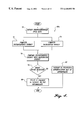
- FIG. 10 is a flow diagram illustrating another pulse detection process performed by a defibrillator as shown in FIG. 2 that incorporates features of the pulse detection processes shown in FIGS. 4, 6 and 9 ;
- FIG. 11 is a flow diagram of a protocol implemented by a defibrillator as shown in FIG. 2 that incorporates a pulse detection process provided by the present invention.
- the present invention may be implemented in a variety of applications, the present invention is particularly suited for use in a defibrillator, such as the defibrillator 10 shown in FIG. 2 .
- the defibrillator 10 is shown connected to a patient 40 via defibrillation electrodes 12 and 14 placed on the skin of the patient 40 .
- the defibrillator 10 uses the defibrillation electrodes 12 and 14 to deliver defibrillation pulses to the patient 40 .
- the defibrillator 10 may also use the electrodes 12 and 14 to obtain ECG signals from the patient 40 .
- FIG. 2 also illustrates sensing devices 16 and 18 placed on the patient 40 .
- the sensing devices 16 and 18 are configured to detect a physiological signal in the patient, preferably acoustical energy from heart sounds produced in the patient 40 .
- the acoustical energy is converted by the defibrillator 10 into digital phonocardiogram (PCG) data.
- the sensing devices 16 and 18 may be integrated into or attached to the back of the electrodes 12 and 14 .
- the sensing devices 16 and 18 may be embodied in flaps 20 and 22 that are connected to the electrodes 12 and 14 .
- the sensing devices 16 and 18 may be attached to the patient 40 by separate wires (not shown).
- the sensing devices 16 and 18 are comprised of piezoelectric transducers.
- the sensing devices 16 and 18 may alternatively be comprised of other acoustic sensors known in the art, such as electronic microphones used in stethoscopes. Transducers and microphones suitable for use in the present invention for detecting heart sounds are described, for example, in U.S. Pat. Nos. 4,446,873 and 5,825,895.
- FIG. 3 illustrates in more detail the major components of the defibrillator 10 shown in FIG. 2 .
- a patient's heart sounds are sensed by PCG/defibrillation electrodes 30 (e.g., a combination of electrodes 12 and 14 with sensing devices 16 and 18 , as described above in reference to FIG. 2) and converted to PCG signals in a conventional manner.
- the PCG/defibrillation electrodes 30 provide the PCG signals to a signal amplifier 32 that amplifies the PCG signals to a level sufficient for the defibrillator 10 to further analyze the PCG signals.
- Alternative embodiments of the defibrillator 10 may include additional signal amplification or signal filtering to adapt the defibrillator 10 for use in particular environments.
- the signal amplifier 32 provides the amplified PCG signals to an anti-aliasing filter 34 .
- the anti-aliasing filter 34 is designed to reduce aliasing introduced in the PCG signals by the analog-to-digital (A/D) converter 36 .
- the bandwidth of the anti-aliasing filter 34 depends, in part, on the sampling rate of the A/D converter 36 .
- Anti-aliasing filters and A/D converters are well-known in the art and are readily available in off-the-shelf devices.
- the A/D converter 36 converts the PCG signals into digitized PCG data and provides the PCG data to a processing unit 38 for evaluation.
- the processing unit 38 evaluates the PCG data using a pulse detection process described below in more detail.
- the processing unit 38 is preferably comprised of a computer processor that operates in accordance with programmed instructions 42 stored in a memory 40 that implement the pulse detection process.
- the processing unit 38 also stores the PCG data in the memory 40 .
- the memory 40 may be comprised of any type of storage medium including, for example, a volatile memory such as a random access memory (RAM), a non-volatile static memory, or a magnetic or optical storage medium (e.g., tape or hard drive).
- the processing unit 38 may report the results of the pulse detection process to the operator of the defibrillator 10 via a display 48 .
- the display 48 may include, for example, lights, audible signals, a printer, or a display screen (e.g., LCD or AMLCD).
- the processing unit 38 may also receive input from the operator of the defibrillator 10 via an input device 46 .
- the input device 46 may include one or more keys, switches, buttons, or other types of user input devices.
- the defibrillator 10 may also use the PCG/defibrillation electrodes 30 to sense the patient's electrocardiogram (ECG) signals.
- ECG electrocardiogram
- the signals obtained from the patient are amplified and filtered by the signal amplifier 32 and further filtered by the anti-aliasing filter 34 in a conventional manner.
- the A/D converter 36 converts the ECG signals into digitized ECG data and provides the ECG data to the processing unit 38 for evaluation.
- the processing unit 38 evaluates the ECG signals in accordance with programmed instructions 44 stored in the memory 40 that carry out an ECG evaluation process to determine whether a defibrillation shock should be provided.
- a suitable method for determining whether to apply a defibrillation shock is described in U.S. Pat. No. 4,610,254, which is assigned to the assignee of the present invention and incorporated by reference herein. If the processing unit 38 determines that delivery of a defibrillation pulse is appropriate, the processing unit 38 instructs a defibrillation pulse generator 50 to prepare to deliver a defibrillation pulse to the patient. In that regard, the defibrillation pulse generator 50 charges one or more defibrillation capacitors in the defibrillator 10 .
- the processing unit 38 advises the operator (e.g., via the display 48 ) that the defibrillator 10 is ready to deliver the defibrillation pulse. Preferably, the processing unit 38 asks the operator to initiate the delivery of the defibrillation pulse.
- the processing unit 38 instructs the defibrillation pulse generator 50 to discharge through the patient the energy stored in the defibrillation capacitors (via the PCG/defibrillation electrodes 30 ).
- the processing unit 38 may cause the defibrillation pulse generator 50 to automatically deliver the defibrillation pulse when specified conditions (e.g., expiration of a predetermined period of time, acceptable measured patient impedance, etc.) are met.
- FIG. 3 illustrates the major components of the defibrillator 10
- the defibrillator 10 may contain more components than those shown in FIG. 3 .
- the disclosure of a preferred embodiment of the defibrillator 10 does not require that all of these general conventional components be shown.
- the invention may be implemented in a cardiac monitor having essentially the same components as the defibrillator 10 shown in FIG. 3, except that the cardiac monitor does not have the components necessary for delivering a defibrillation pulse.
- the programmed instructions 42 may be encoded in hardware as an alternative to software instructions stored in the memory 40 .
- the pulse detection process conducted by the processing unit 38 analyzes the patient's PCG data to determine whether heart sounds S 1 and/or S 2 are present.
- the presence of heart sounds S 1 and/or S 2 are used as a surrogate to indicate the presence of a cardiac pulse in the patient.
- the pulse detection process detects heart sounds in the patient's PCG, the pulse detection process determines that a pulse is present in the patient.
- FIG. 4 illustrates one version of the pulse detection process 60 a using a temporal energy analysis conducted in accordance with the present invention.
- the pulse detection process 60 a begins in a block 70 by obtaining PCG data from a patient.
- PCG signals obtained by PCG sensing devices e.g., sensing devices 16 and 18 in FIG. 2 placed on the patient are converted into digital PCG data.
- the pulse detection process 60 a evaluates the PCG data for at least one feature indicative of the presence of a heart sound.
- the pulse detection process 60 a preferably calculates estimates of the instantaneous energy and background energy in the PCG data. As shown in FIG. 4, the estimated instantaneous energy may be calculated in block 72 simultaneously with the calculation of estimated background energy in block 74 . Alternatively, the calculation of estimated instantaneous energy in block 72 may be performed prior to or after the calculation of estimated background energy in block 74 .
- the estimated instantaneous energy is calculated in block 72 , preferably using a set of PCG data obtained from the patient during a predetermined time window.
- the time window is 20 milliseconds in length, though a longer, shorter, or slightly shifted time window may be used for estimating the instantaneous energy.
- the estimated instantaneous energy is preferably calculated by squaring and summing each of the PCG data values in the predetermined time window.
- the estimated background energy is calculated in block 74 , preferably using a set of PCG data obtained in an earlier predetermined time window.
- the estimated background energy is calculated using PCG data in a 20 millisecond time window commencing 50 milliseconds prior to the current time.
- the PCG data within the earlier time window are also preferably squared and summed to produce the estimated background energy.
- the estimated instantaneous energy and background energy are next compared in a block 76 to determine a relative change in energy in the PCG data.
- the relative change in energy is used by the pulse detection process 60 a as a feature indicative of the presence of a heart sound. If the relative change in energy between the estimated instantaneous energy and the estimated background energy exceeds a predetermined threshold, the pulse detection process 60 a determines that a heart sound was detected.
- the present invention uses the detection of a heart sound as an indication of the presence of a cardiac pulse in the patient.
- the pulse detection process 60 a proceeds to a block 80 and reports the presence of a cardiac pulse in the patient (thus indicating that defibrillation therapy for the patient is not advised). Otherwise, if a heart sound is not detected, the pulse detection process 60 a determines in a block 82 that the patient is pulseless and that defibrillation therapy may be appropriate.
- a defibrillator 10 implementing the pulse detection process 60 a may then proceed to determine whether defibrillation therapy is appropriate, e.g., by obtaining and processing ECG data from the patient as described in U.S. Pat. No. 4,610,254, referenced earlier.
- the pulse detection process 60 a may be repeated over a specified interval of time or for a specified number of repetitions to produce a series of determinations of whether a heart sound is present in the patient.
- the time windows for computing the estimated instantaneous energy and background energy are shifted to correspond with each instance of time in which the pulse detection process 60 a is performed.
- the pulse detection process 60 a may require a specified number of heart sound detections before determining that a cardiac pulse is present in the patient.
- FIGS. 5A-5D illustrate a representative example of the processing performed by the pulse detection process 60 a .
- FIG. 5A is a graph showing a PCG waveform 84 of raw PCG data as collected in block 70 (FIG. 4) from a patient.
- the PCG data may be filtered to reduce noise and other signal contaminants.
- a filtered version of the PCG waveform 86 is shown in FIG. 5 B.
- FIG. 5C illustrates a waveform 88 depicting the estimated instantaneous energy in the PCG as calculated in block 72 of the pulse detection process 60 a .
- the waveform 90 depicts the estimated background energy as calculated in block 74 of the pulse detection process 60 a . Because the calculation of background energy 90 uses PCG data obtained in an earlier time window than the PCG data used to calculate instantaneous energy 88 , the rise and fall of the background energy waveform 90 follows the rise and fall of the instantaneous energy waveform 88 .
- the comparison performed in block 76 of the pulse detection process 60 a may produce a result as illustrated in FIG. 5 D.
- the comparison performed in block 76 returns a “1” (signifying the detection of a heart sound), as noted by reference numeral 92 .
- the predetermined threshold may be adjusted to achieve a desired sensitivity and specificity of detection.
- the comparison performed in block 76 returns a “0”, as noted by reference number 94 , signifying that a heart sound was not detected.
- FIG. 6 illustrates another version of the pulse detection process 60 b .
- the version 60 b analyzes PCG data to detect heart sounds in a patient. If heart sounds are detected, the pulse detection process 60 b determines that a pulse is present in the patient.
- the version 60 b of the pulse detection process focuses on a spectral energy analysis of the PCG data (as compared to the temporal energy analysis performed in the version 60 a ).
- the pulse detection process 60 b begins in a block 100 by obtaining PCG data from the patient in a manner as discussed above with respect to block 70 (FIG. 4 ).
- the PCG data is preferably analyzed to identify a set of PCG data that likely contains an S 1 or S 2 heart sound.
- an S 1 or S 2 heart sound candidate may be identified by using the energy comparison discussed in block 76 of the pulse detection process 60 a .
- the energy comparison indicates that a potential S 1 or S 2 candidate has been detected.
- a set of PCG data containing a heart sound may be identified by evaluating the patient's ECG data for the occurrence of an R-wave.
- the timing of an S 1 or S 2 heart sound in relation to an R-wave is generally known in the art and may be used to predict the timing of a heart sound candidate in the PCG data.
- the pulse detection process 60 b computes an energy spectrum of the heart sound candidate, preferably using a maximum entropy method.
- a maximum entropy method (“MEM spectrum”) is well-known in the art. See, e.g., Modern Spectral Estimation: Theory and Application , by Stephen M. Kay, published by Prentice Hall of Englewood Cliffs, N.J., p. 182, incorporated herein by reference.
- An MEM spectrum typically appears much smoother than an energy spectrum produced by Fourier transform techniques.
- FIG. 7 illustrates a representative MEM spectrum 120 for an interval of PCG data containing an S 1 heart sound.
- FIG. 7 also illustrates a representative MEM spectrum 130 for a set of PCG data containing an S 2 heart sound.
- the MEM spectrum 120 includes a number of peak energy values, including the first two peak values 122 and 124 .
- the MEM spectrum 130 includes a number of peak energy values, including the first two peak values 132 and 134 .
- the MEM spectrum 120 or 130 whichever is used, may be normalized by removing the baseline (DC) energy value across the MEM spectrum.
- the frequency of a peak energy value in the energy spectrum is used as a feature indicative of the presence of a heart sound, and is evaluated against a predetermined threshold frequency value to decide whether a heart sound is detected.
- the pulse detection process 60 b in FIG. 6 evaluates the second peak energy value occurring in the energy spectrum measured from DC, e.g., the second peak value 124 in the MEM spectrum 120 , or the second peak value 134 in the MEM spectrum 130 .
- the pulse detection process 60 b evaluates the energy values in the MEM spectrum to determine the frequency of the second peak in the MEM spectrum. For example, if the pulse detection process 60 b evaluates MEM spectrum 120 , the frequency of the second peak 124 is determined. A similar analysis applied to the MEM spectrum 130 determines the frequency of the second peak 134 .
- the frequency of the second peak 124 or 134 is compared with a predetermined threshold frequency to decide whether a heart sound is detected. For example, if the frequency of the second peak 124 or 134 is less than or equal to a threshold frequency, e.g., 100 Hz, the pulse detection process 60 b determines that a heart sound was detected.
- a threshold frequency e.g. 100 Hz
- Alternative embodiments of the invention may use values other than 100 Hz for the predetermined threshold frequency.
- the pulse detection process 60 b proceeds from decision block 110 to a block 112 and determines that a pulse is present in the patient, thus advising against application of a defibrillation pulse. If, in decision block 110 , a heart sound was not detected, the pulse detection process 60 b determines in a block 114 that the patient is pulseless and that defibrillation may be appropriate for the patient. In that case, further signal processing of ECG data obtained from the patient is preferably performed to determine the applicability of defibrillation therapy, e.g., as described in U.S. Pat. No. 4,610,254, referenced earlier.
- FIG. 8A is a graph depicting a PCG waveform 140 of raw PCG data obtained from a patient in a manner as discussed above in regard to block 100 (FIG. 6 ).
- the PCG waveform 140 may be filtered to reduce noise and other signal contaminants (e.g., as described earlier in reference to FIG. 5 B).
- an MEM spectrum of the data in the PCG waveform 140 is computed for a number of instances in time, and the frequency of the second peak of each MEM spectrum is identified, as shown by the circles in FIG. 8B, without regard to whether the selected instance of time corresponds with a heart sound candidate.
- the PCG data first be screened for heart sound candidates.
- the circles enclosing an “x” identify the MEM spectra that, for this example, have a second peak located at or below a threshold frequency of 100 Hz. Note that, for the most part, the circles with an “x” in FIG. 8B correspond in time with the heart sounds evident in the PCG waveform 140 shown in FIG. 8 A. For each circled “x,” the pulse detection process 60 b decides that a heart sound, and thus a pulse, is present in the patient.
- FIG. 9 illustrates another version 60 c of the pulse detection process that also uses an MEM spectrum as calculated in block 104 of the version 60 b .
- the version 60 c analyzes the energy value of the second peak in the MEM spectrum.
- the version 60 c of the pulse detection process begins in a block 150 by obtaining PCG data from the patient in a manner as discussed earlier with respect to block 70 (FIG. 4 ).
- the PCG data is analyzed in a block 152 to identify PCG data corresponding to the time when a heart sound S 1 or S 2 likely occurred.
- the analysis performed in block 152 may include an energy comparison process or ECG analysis as described earlier with respect to block 102 of pulse detection process 60 b (FIG. 6 ).
- An MEM spectrum of the heart sound candidate is then computed in a block 154 in a manner as discussed earlier with respect to block 104 (FIG. 6 ).
- the pulse detection process 60 c evaluates the energy values in the MEM spectrum to locate the second peak value in the spectrum.
- the energy value of the second peak determined in a block 158 , is used as a feature indicative of the presence of a heart sound, and is compared in a block 160 with a threshold energy to decide whether a heart sound was detected. If the energy value of the second peak exceeds the threshold energy, the pulse detection process 60 c determines in a decision block 162 that a heart sound was detected.
- the pulse detection process 60 c determines in a block 164 that a cardiac pulse is present in the patient and advises against providing defibrillation therapy to the patient. On the other hand, if a heart sound was not detected in decision block 162 , the pulse detection process 60 c determines in a block 166 that the patient is pulseless and advises that defibrillation therapy may be appropriate for the patient.
- An analysis of ECG data may be used to determine the applicability of defibrillation therapy.
- FIGS. 8A and 8C illustrate one example of the processing performed by the pulse detection process 60 c .
- FIG. 8A illustrates a PCG waveform 140 of raw PCG data obtained from a patient from which an MEM spectrum is computed for a number of instances in time. For each instance in time, the energy value of the second peak in the MEM spectrum is identified, as depicted by the circles in FIG. 8 C.
- the circles enclosing an “x” are the MEM spectra with a second peak having an energy value above a selected threshold energy, e.g., 0 dB. While a threshold value of 0 dB is used in this specific example, other embodiments of the invention may use different threshold values.
- the circles with an “x” in FIG. 8C generally correspond in time with the heart sounds evident in the PCG waveform 140 shown in FIG. 8 A.
- the pulse detection process 60 c decides that a heart sound, and hence a cardiac pulse, is present in the patient.
- noise in the PCG data may cause a false detection of a heart sound when using one of the versions 60 a , 60 b , or 60 c of the pulse detection process described above. See, e.g., the two circled x's in FIGS. 8B and 8C immediately following the time reference of 0.6 seconds, which do not appear to correspond with heart sounds evident in FIG. 8 A.
- the versions 60 a , 60 b , and 60 c of the pulse detection process may be combined in one or more ways to produce a version of the pulse detection process that improves the specificity of the heart sound detection.
- FIG. 10 illustrates a version 60 d of the pulse detection process that combines features of the versions 60 a , 60 b , and 60 c of the pulse detection process.
- the pulse detection process 60 d begins in a block 170 by obtaining PCG data from a patient, e.g., in a manner as described earlier with respect to block 70 of pulse detection process 60 a (FIG. 4 ).
- estimates of the instantaneous energy and the background energy in the PCG data are computed in blocks 172 and 174 , e.g., in a manner as described earlier with respect to blocks 72 and 74 .
- the estimated instantaneous and background energy values are then compared in a block 176 , e.g., as described earlier with respect to block 76 , to produce a first detection statistic, or feature, indicative of the presence of a heart sound.
- the first detection statistic produced in block 176 is provided to a classifier in block 186 that evaluates detection statistics to determine whether a heart sound was present.
- a classifier in block 186 that evaluates detection statistics to determine whether a heart sound was present.
- the instantaneous and background energies computed in blocks 172 and 174 may also be directly provided as separate detection statistics to a multi-dimensional classifier in block 186 for joint classification with any other detection statistics provided to a multi-dimensional classifier (i.e., eliminating the comparison performed in block 176 ).
- the PCG data obtained in block 170 is also used in identifying a heart sound candidate and computing an MEM spectrum in block 178 , in a manner as described earlier with respect to blocks 102 and 104 of pulse detection process 60 b (FIG. 6 ).
- the pulse detection process 60 d determines in a block 180 the location of the second peak in the MEM spectrum.
- the frequency of the second peak is determined in a block 182 and provided as a second detection statistic, or feature, to the classifier in block 186 .
- the second detection statistic may be produced by comparing the frequency of the second peak with a threshold frequency, e.g., in a manner as described earlier with respect to block 108 (FIG. 6 ).
- the pulse detection process 60 d also determines the energy value at the second peak and provides the energy value as a third detection statistic, or feature, to the classifier in block 186 .
- the second peak energy may alternatively be compared with a threshold energy, e.g., in a manner as described earlier with respect to block 160 (FIG. 9 ), to produce the third detection statistic.
- the classifier in block 186 jointly classifies the first, second, and third detection statistics using a multi-dimensional classifier to determine whether a heart sound, and hence a pulse, is present in the patient.
- Techniques for constructing multidimensional classifiers are well-known in the art.
- For an expanded description of a classifier suitable for use in the present invention see, e.g., R. Duda and P. Hart, Pattern Classification and Scene Analysis , published by John Wiley & Sons, New York, and incorporated herein by reference.
- the classifier in block 186 may also use a voting scheme to determine whether a pulse is present in the patient. For example, if any of the first, second, or third detection statistics indicates the detection of a heart sound (i.e., the instantaneous energy exceeded the background energy by a threshold value, the frequency of the second peak was equal to or less than a threshold frequency, or the energy of the second peak exceeded a threshold energy), the classifier determines that a pulse is present in the patient. Alternatively, the classifier in block 186 may determine that a pulse is present by finding that a combination of the first, second, and third detection statistics indicates the presence of a heart sound (e.g., a positive indication from the first detection statistic combined with a positive indication from the second or third detection statistics, etc.). The classifier in block 186 may also weight the first, second, or third detection statistics to emphasize one detection statistic over another in deciding whether a heart sound was detected.
- a voting scheme to determine whether a pulse is present in the patient. For example, if any of the
- the pulse detection process 60 d determines in a block 190 that a pulse is present in the patient and advises the operator of the defibrillator to not defibrillate the patient. Otherwise, if a heart sound was not detected in decision block 188 , the pulse detection process 60 d determines in a block 192 that the patient is pulseless and that defibrillation therapy may be appropriate.
- An analysis of ECG data as described earlier in reference to U.S. Pat. No. 4,610,254, may be used to determine if defibrillation therapy is appropriate.
- the pulse detection process of the invention may be used as part of a protocol in a defibrillator for determining whether to provide defibrillation therapy to the patient.
- FIG. 11 illustrates one implementation of a pulse detection/defibrillation process 200 , preferably for use in an automated external defibrillator (AED) capable of providing a defibrillation pulse if a patient is determined to be in cardiac arrest.
- AED automated external defibrillator
- the process 200 proceeds to a block 202 where the AED initializes its circuits. The AED's PCG/defibrillation electrodes are placed on the patient.
- the process 200 proceeds to a block 204 where the AED performs an analysis of the patient in which the AED obtains selected parameters such as PCG data and ECG data from the patient. During the analysis performed in block 204 , the AED reports “Analyzing now . . . stand clear” to the operator of the AED.
- the process 200 jumps to a block 206 where the AED instructs the operator to “Connect electrodes.” When the AED senses that the electrodes are connected, the process 200 returns to the analysis in block 204 . Likewise, if the AED finds itself in any other state where the electrodes are not connected, as represented by block 208 , the process 200 jumps to block 206 where it instructs the operator to connect the electrodes.
- the process 200 proceeds to a block 210 where the AED reports to the operator of the AED “Motion detected . . . stop motion.” If the patient is moved during the analysis process 204 , the data obtained during the analysis is more likely to be contaminated with noise and other signal contaminants.
- motion on the part of the patient is detected by an impedance channel that evaluates the impedance measured between the PCG/defibrillation electrodes placed on the patient. If the measured impedance fluctuates outside of a predetermined range, the AED determines that the patient is moving and directs the process 200 to proceed to block 210 . When the motion ceases, the process 200 returns to the analysis in block 204 .
- the pulse detection performed in block 212 may be any one or combination of the versions 60 a , 60 b , 60 c , or 60 d of the pulse detection process described above, and may involve an analysis of a physiological signal sensed in the patient other than acoustical energy (PCG data).
- PCG data acoustical energy
- a pulse is not detected in the patient, the process 200 proceeds to a decision block 214 where it determines whether the patient has a shockable cardiac rhythm (e.g., ventricular fibrillation (VF) or ventricular tachycardia (VT)) or a non-shockable cardiac rhythm (such as asystole and bradycardia).
- a shockable cardiac rhythm e.g., ventricular fibrillation (VF) or ventricular tachycardia (VT)
- VT ventricular tachycardia
- non-shockable cardiac rhythm such as asystole and bradycardia
- the process 200 proceeds to a block 216 where the AED prepares to deliver a defibrillation pulse to the patient.
- an energy storage device such as a capacitor bank, within the AED is charged to a specified level.
- the AED reports “Shock advised” to the operator of the AED.
- the process 200 proceeds to a block 218 where the AED is ready to deliver the defibrillation pulse.
- the operator of the AED is advised “Stand clear . . . push to shock.”
- the process 200 proceeds to block 220 and delivers the defibrillation shock to the patient.
- the AED preferably records in memory that it delivered a defibrillation pulse to the patient. If the present pulse delivery is the first or second defibrillation shock delivered to the patient, the process 200 returns to block 204 where the patient undergoes another analysis.
- the process 200 proceeds to a block 222 where the AED advises the operator to commence providing CPR therapy to the patient, e.g., by using the message “No shock advised . . . start CPR.”
- the process 200 proceeds to block 222 and advises the operator to provide CPR therapy. Again, at this point, the AED reports “No shock advised . . . start CPR” to the operator. Preferably, the prompt to provide CPR is provided for a defined period of time. When the period of time for CPR is finished, the process 200 returns to block 204 and performs another analysis on the patient.
- the process 200 proceeds to a decision block 224 where it determines whether the patient is breathing.
- the AED may use any one of a number of conventional means for automatically determining whether a patient is breathing. Such means include attaching a sample tube by the patient's mouth to analyze fluctuations in carbon dioxide exhaled by the patient. Other means include observing changes in impedance of the patient that are indicative of a change in volume in the patient's lungs. Other means also include stretching a band with a strain gauge around the patient's chest to observe expansion of the patient's chest due to breathing. Alternatively, if automatic means for detecting breathing in the patient are not available, the AED may ask the operator of the AED to input information (e.g., by pressing a button) to indicate whether the patient is breathing.
- the process 200 determines that the patient is not breathing, the process 200 proceeds to a block 226 where the operator of the AED is advised to commence rescue breathing. In that regard, the AED reports to the operator “Pulse detected . . . start rescue breathing.” The AED also continues to monitor the patient's cardiac pulse and returns to block 204 if a cardiac pulse is no longer detected. If, at any point during the provision of rescue breathing, the AED detects that the patient is breathing on his own, the process 200 proceeds to a block 228 where the AED monitors the patient for a continued presence of breathing and a cardiac pulse.
- the process 200 proceeds to block 228 where the AED monitors the status of the patient. In that regard, the AED reports “Pulse and breathing detected . . . monitoring patient.” If, at any time during the monitoring of the patient the process 200 determines that the patient is not breathing, the process 200 proceeds to block 226 where the operator of the AED is advised to commence rescue breathing. If a cardiac pulse is no longer detected in the patient, the process 200 proceeds from block 228 to block 204 to commence a new analysis of the patient.
- the AED may assess whether CPR is being administered to the patient. If the AED finds that CPR is being performed, the AED may prompt the operator to cease providing CPR. If, during the CPR period of block 222 , the AED determines that CPR is not being administered to the patient, the AED may remind the operator to provide CPR therapy to the patient.
- One method for determining whether CPR is being administered is to monitor patient impedance to observe patterns of impedance fluctuation in the patient that are indicative of CPR.
- an analysis of ECG data may be combined with an analysis of PCG data to determine the presence of a cardiac pulse in the patient.
- detecting a QRS complex in the ECG data preceding the time that a heart sound occurs may serve to confirm the detection of the heart sound.
- detecting a QRS complex in the ECG data may be used to identify PCG data for use in the heart sound detection process, since a heart sound is expected to occur after the occurrence of a QRS complex if a cardiac pulse is present in the patient. This aspect of the invention is helpful in identifying a heart sound candidate in the PCG data. It is also helpful in identifying whether the patient is in a state of pulseless electrical activity.
- the patient is in a state of pulseless electrical activity that may be reported to the operator of the device (e.g., along with the CPR advisory in block 222 of the pulse detection/defibrillation process 200 shown in FIG. 11 ).
- the pulse detection processes described in detail herein primarily use an analysis of PCG data to determine the presence of a cardiac pulse
- the pulse detection processes may analyze data obtained from other physiological signals sensed in the patient for features indicative of the presence of a cardiac pulse. For instance, variations in the patient's transthoracic impedance may be associated with the discharge of blood from the heart. By monitoring characteristic variations in the patient's transthoracic impedance, the pulse detection process may monitor the patient's cardiac output, and hence determine the presence of a cardiac pulse. Ultrasound signals from Doppler ultrasound probes placed on the patient may also be used to monitor and quantify blood flow in the patient, and hence determine the presence of a cardiac pulse.
Abstract
Description
Claims (62)
Priority Applications (19)
| Application Number | Priority Date | Filing Date | Title |
|---|---|---|---|
| US09/410,198 US6440082B1 (en) | 1999-09-30 | 1999-09-30 | Method and apparatus for using heart sounds to determine the presence of a pulse |
| JP2001526103A JP2003510123A (en) | 1999-09-30 | 2000-09-25 | Method and apparatus for using heart sounds to determine the presence of a pulse |
| DE60028984T DE60028984T2 (en) | 1999-09-30 | 2000-09-25 | DEVICE FOR USING HEARTS TO DETERMINE THE PRESENCE OF A PULSE |
| EP00977256A EP1216000B1 (en) | 1999-09-30 | 2000-09-25 | Apparatus for using heart sounds to determine the presence of a pulse |
| PCT/US2000/040980 WO2001022885A1 (en) | 1999-09-30 | 2000-09-25 | Method and apparatus for using heart sounds to determine the presence of a pulse |
| US10/229,321 US20040039419A1 (en) | 1999-09-30 | 2002-08-26 | Apparatus, software, and methods for cardiac pulse detection using a piezoelectric sensor |
| US10/229,320 US20030060723A1 (en) | 1999-09-30 | 2002-08-26 | Pulse detection apparatus, software, and methods using patient physiological signals |
| US11/167,247 US7917209B2 (en) | 1999-09-30 | 2005-06-27 | Pulse detection apparatus, software, and methods using patient physiological signals |
| US11/187,616 US8160703B2 (en) | 1999-09-30 | 2005-07-22 | Apparatus, software, and methods for cardiac pulse detection using a piezoelectric sensor |
| US11/737,703 US8092392B2 (en) | 1999-09-30 | 2007-04-19 | Pulse detection method and apparatus using patient impedance |
| US13/032,250 US8239024B2 (en) | 1999-09-30 | 2011-02-22 | Pulse detection apparatus, software, and methods using patient physiological signals |
| US13/249,081 US20120022339A1 (en) | 1999-09-30 | 2011-09-29 | Pulse detection apparatus, software, and methods using patient physiological signals |
| US13/267,783 US8532766B2 (en) | 1999-09-30 | 2011-10-06 | Pulse detection apparatus, software, and methods using patient physiological signals |
| US13/267,769 US20120029368A1 (en) | 1999-09-30 | 2011-10-06 | Pulse detection apparatus, software, and methods using patient physiological signals |
| US13/272,733 US20120029584A1 (en) | 1999-09-30 | 2011-10-13 | Pulse detection apparatus, software, and methods using patient physiological signals |
| US13/272,703 US20120035678A1 (en) | 1999-09-30 | 2011-10-13 | Pulse detection apparatus, software, and methods using patient physiological signals |
| US13/569,147 US8744577B2 (en) | 1999-09-30 | 2012-08-07 | Pulse detection apparatus, software, and methods using patient physiological signals |
| US13/970,522 US9248306B2 (en) | 1999-09-30 | 2013-08-19 | Pulse detection apparatus, software, and methods using patient physiological signals |
| US14/976,976 US9981142B2 (en) | 1999-09-30 | 2015-12-21 | Pulse detection apparatus, software, and methods using patient physiological signals |
Applications Claiming Priority (1)
| Application Number | Priority Date | Filing Date | Title |
|---|---|---|---|
| US09/410,198 US6440082B1 (en) | 1999-09-30 | 1999-09-30 | Method and apparatus for using heart sounds to determine the presence of a pulse |
Related Parent Applications (1)
| Application Number | Title | Priority Date | Filing Date |
|---|---|---|---|
| US10/013,941 Continuation-In-Part US20030109790A1 (en) | 1999-09-30 | 2001-12-06 | Pulse detection method and apparatus using patient impedance |
Related Child Applications (2)
| Application Number | Title | Priority Date | Filing Date |
|---|---|---|---|
| US10/013,941 Continuation-In-Part US20030109790A1 (en) | 1999-09-30 | 2001-12-06 | Pulse detection method and apparatus using patient impedance |
| US10/229,320 Continuation-In-Part US20030060723A1 (en) | 1999-09-30 | 2002-08-26 | Pulse detection apparatus, software, and methods using patient physiological signals |
Publications (1)
| Publication Number | Publication Date |
|---|---|
| US6440082B1 true US6440082B1 (en) | 2002-08-27 |
Family
ID=23623685
Family Applications (10)
| Application Number | Title | Priority Date | Filing Date |
|---|---|---|---|
| US09/410,198 Expired - Lifetime US6440082B1 (en) | 1999-09-30 | 1999-09-30 | Method and apparatus for using heart sounds to determine the presence of a pulse |
| US10/229,320 Abandoned US20030060723A1 (en) | 1999-09-30 | 2002-08-26 | Pulse detection apparatus, software, and methods using patient physiological signals |
| US11/167,247 Expired - Fee Related US7917209B2 (en) | 1999-09-30 | 2005-06-27 | Pulse detection apparatus, software, and methods using patient physiological signals |
| US13/032,250 Expired - Fee Related US8239024B2 (en) | 1999-09-30 | 2011-02-22 | Pulse detection apparatus, software, and methods using patient physiological signals |
| US13/249,081 Abandoned US20120022339A1 (en) | 1999-09-30 | 2011-09-29 | Pulse detection apparatus, software, and methods using patient physiological signals |
| US13/267,769 Abandoned US20120029368A1 (en) | 1999-09-30 | 2011-10-06 | Pulse detection apparatus, software, and methods using patient physiological signals |
| US13/267,783 Expired - Fee Related US8532766B2 (en) | 1999-09-30 | 2011-10-06 | Pulse detection apparatus, software, and methods using patient physiological signals |
| US13/272,703 Abandoned US20120035678A1 (en) | 1999-09-30 | 2011-10-13 | Pulse detection apparatus, software, and methods using patient physiological signals |
| US13/272,733 Abandoned US20120029584A1 (en) | 1999-09-30 | 2011-10-13 | Pulse detection apparatus, software, and methods using patient physiological signals |
| US13/569,147 Expired - Fee Related US8744577B2 (en) | 1999-09-30 | 2012-08-07 | Pulse detection apparatus, software, and methods using patient physiological signals |
Family Applications After (9)
| Application Number | Title | Priority Date | Filing Date |
|---|---|---|---|
| US10/229,320 Abandoned US20030060723A1 (en) | 1999-09-30 | 2002-08-26 | Pulse detection apparatus, software, and methods using patient physiological signals |
| US11/167,247 Expired - Fee Related US7917209B2 (en) | 1999-09-30 | 2005-06-27 | Pulse detection apparatus, software, and methods using patient physiological signals |
| US13/032,250 Expired - Fee Related US8239024B2 (en) | 1999-09-30 | 2011-02-22 | Pulse detection apparatus, software, and methods using patient physiological signals |
| US13/249,081 Abandoned US20120022339A1 (en) | 1999-09-30 | 2011-09-29 | Pulse detection apparatus, software, and methods using patient physiological signals |
| US13/267,769 Abandoned US20120029368A1 (en) | 1999-09-30 | 2011-10-06 | Pulse detection apparatus, software, and methods using patient physiological signals |
| US13/267,783 Expired - Fee Related US8532766B2 (en) | 1999-09-30 | 2011-10-06 | Pulse detection apparatus, software, and methods using patient physiological signals |
| US13/272,703 Abandoned US20120035678A1 (en) | 1999-09-30 | 2011-10-13 | Pulse detection apparatus, software, and methods using patient physiological signals |
| US13/272,733 Abandoned US20120029584A1 (en) | 1999-09-30 | 2011-10-13 | Pulse detection apparatus, software, and methods using patient physiological signals |
| US13/569,147 Expired - Fee Related US8744577B2 (en) | 1999-09-30 | 2012-08-07 | Pulse detection apparatus, software, and methods using patient physiological signals |
Country Status (5)
| Country | Link |
|---|---|
| US (10) | US6440082B1 (en) |
| EP (1) | EP1216000B1 (en) |
| JP (1) | JP2003510123A (en) |
| DE (1) | DE60028984T2 (en) |
| WO (1) | WO2001022885A1 (en) |
Cited By (77)
| Publication number | Priority date | Publication date | Assignee | Title |
|---|---|---|---|---|
| US20020032383A1 (en) * | 2000-07-21 | 2002-03-14 | Weil Max Harry | Cardiac/respiratory arrest detector |
| US20020156389A1 (en) * | 2001-02-28 | 2002-10-24 | Cardiac Pacemakers, Inc. | Cardiac rhythm management patient report |
| US20020165585A1 (en) * | 2001-05-01 | 2002-11-07 | Dupelle Michael R. | Pulse sensors |
| US20030018276A1 (en) * | 2000-10-06 | 2003-01-23 | Mansy Hansen A. | Acoustic detection of endotracheal tube location |
| US20030060723A1 (en) * | 1999-09-30 | 2003-03-27 | Medtronic Physio-Control Manufacturing Corp. | Pulse detection apparatus, software, and methods using patient physiological signals |
| US6575914B2 (en) * | 2001-05-18 | 2003-06-10 | Koninklijke Philips Electronics N.V. | Integrated cardiac resuscitation system with ability to detect perfusion |
| US20030109790A1 (en) * | 2001-12-06 | 2003-06-12 | Medtronic Physio-Control Manufacturing Corp. | Pulse detection method and apparatus using patient impedance |
| US20030144699A1 (en) * | 2002-01-25 | 2003-07-31 | Koninkilijke Philips Electronics N.V. | System and method for determining a condition of a patient |
| US20040039420A1 (en) * | 2002-08-26 | 2004-02-26 | Medtronic Physio-Control Manufacturing Corp. | Apparatus, software, and methods for cardiac pulse detection using accelerometer data |
| US20040039419A1 (en) * | 1999-09-30 | 2004-02-26 | Stickney Ronald E. | Apparatus, software, and methods for cardiac pulse detection using a piezoelectric sensor |
| US20040116969A1 (en) * | 2002-08-26 | 2004-06-17 | Owen James M. | Pulse detection using patient physiological signals |
| US20040193065A1 (en) * | 2003-03-31 | 2004-09-30 | Houben Richard P.M. | Biomedical signal denoising techniques |
| US20040210147A1 (en) * | 2003-04-16 | 2004-10-21 | Houben Richard P.M. | Biomedical signal analysis techniques using wavelets |
| WO2004073787A3 (en) * | 2003-02-18 | 2004-11-25 | Purdue Research Foundation | Apparatus and method for noninvasively detecting the quality of cardiac pumping |
| US20040260188A1 (en) * | 2003-06-17 | 2004-12-23 | The General Hospital Corporation | Automated auscultation system |
| US20050027317A1 (en) * | 2003-01-27 | 2005-02-03 | Langer Alois A. | Defibrillation system for non-medical environments |
| US20050038350A1 (en) * | 2003-04-11 | 2005-02-17 | Apurv Kamath | Biopotential signal source separation using source impedances |
| US20050038360A1 (en) * | 2003-04-23 | 2005-02-17 | Hemchandra Shertukde | Apparatus and method for non-invasive diagnosing of coronary artery disease |
| US20050043763A1 (en) * | 2003-04-23 | 2005-02-24 | Zoll Medical Corporation, A Massachusetts Corporation | Processing pulse signal in conjunction with ECG signal to detect pulse in external defibrillation |
| US20050090755A1 (en) * | 2003-10-22 | 2005-04-28 | Guion Marie A. | Analysis of auscultatory sounds using single value decomposition |
| US20050119708A1 (en) * | 2003-04-11 | 2005-06-02 | Paul Haefner | Subcutaneous cardiac signal discrimination employing non-electrophysiologic signal |
| US20050245974A1 (en) * | 2004-04-30 | 2005-11-03 | Sherman Lawrence D | Methods and devices to guide therapy for ventricular fibrillation based on waveform analysis and survival benefit analysis |
| US20050283082A1 (en) * | 2004-06-21 | 2005-12-22 | Geddes Leslie A | Optical noninvasive vital sign monitor |
| US6984207B1 (en) | 1999-09-14 | 2006-01-10 | Hoana Medical, Inc. | Passive physiological monitoring (P2M) system |
| US20060167385A1 (en) * | 2005-01-24 | 2006-07-27 | 3M Innovative Properties Company | Analysis of auscultatory sounds using voice recognition |
| US20060200033A1 (en) * | 2003-05-12 | 2006-09-07 | Cheetah Medical Inc. C/O Pepper Hamiton | System, method and apparatus for measuring blood flow and blood volume |
| US20060241702A1 (en) * | 2005-04-22 | 2006-10-26 | Gillberg Jeffrey M | Cardiac sensing and detection using subcutaneous ECG signals and heart sounds |
| US20060253159A1 (en) * | 2005-05-05 | 2006-11-09 | Siejko Krzysztof Z | Trending of systolic murmur intensity for monitoring cardiac disease with implantable device |
| US7139609B1 (en) | 2003-01-17 | 2006-11-21 | Pacesetter, Inc. | System and method for monitoring cardiac function via cardiac sounds using an implantable cardiac stimulation device |
| US20060270939A1 (en) * | 2005-05-24 | 2006-11-30 | Cardiac Pacemakers, Inc. | Systems and methods for multi-axis cardiac vibration measurements |
| US20060276849A1 (en) * | 2005-06-01 | 2006-12-07 | Cardiac Pacemakers, Inc. | Sensing rate of change of pressure in the left ventricle with an implanted device |
| US20060287606A1 (en) * | 2005-06-21 | 2006-12-21 | Di-Ann Hong | Method for detecting heart rate and systems thereof |
| WO2007010422A2 (en) * | 2005-07-15 | 2007-01-25 | Koninklijke Philips Electronics N.V. | Apparatus and method for defibrillation with pulse detection using electromagnetic waves |
| US20070055151A1 (en) * | 2005-01-20 | 2007-03-08 | Shertukde Hemchandra M | Apparatus and methods for acoustic diagnosis |
| US20070078491A1 (en) * | 2003-12-24 | 2007-04-05 | Cardiac Pacemakers, Inc. | Third heart sound activity index for heart failure monitoring |
| US7248923B2 (en) | 2003-11-06 | 2007-07-24 | Cardiac Pacemakers, Inc. | Dual-use sensor for rate responsive pacing and heart sound monitoring |
| US20080119749A1 (en) * | 2006-11-20 | 2008-05-22 | Cardiac Pacemakers, Inc. | Respiration-synchronized heart sound trending |
| US20080125820A1 (en) * | 2006-11-29 | 2008-05-29 | Cardiac Pacemakers, Inc. | Adaptive sampling of heart sounds |
| US20080262368A1 (en) * | 2007-04-17 | 2008-10-23 | Cardiac Pacemakers, Inc. | Heart sound tracking system and method |
| US20090018449A1 (en) * | 2006-02-03 | 2009-01-15 | Koninklijke Philips Electronics, N.V. | Ultrasonic Method and Apparatus for Measuring or Detecting Flow Behavior of a Non-Sinusoidal Periodicity |
| US20090018461A1 (en) * | 2003-12-24 | 2009-01-15 | Cardiac Pacemakers, Inc. | Method and apparatus for third heart sound detection |
| US20090112274A1 (en) * | 2004-01-23 | 2009-04-30 | Herbert Kevin J | Defibrillator with remote region on its casing |
| US7559901B2 (en) | 2004-07-28 | 2009-07-14 | Cardiac Pacemakers, Inc. | Determining a patient's posture from mechanical vibrations of the heart |
| US20090270930A1 (en) * | 2008-04-25 | 2009-10-29 | Physio-Control, Inc. | External Defibrillator With Adaptive Protocols |
| US20090312659A1 (en) * | 2005-07-26 | 2009-12-17 | Carlson Gerrard M | Managing preload reserve by tracking the ventricular operating point with heart sounds |
| US20100031959A1 (en) * | 2007-03-07 | 2010-02-11 | Cheetah Medical Ltd. | Method and system for monitoring sleep |
| US7662104B2 (en) | 2005-01-18 | 2010-02-16 | Cardiac Pacemakers, Inc. | Method for correction of posture dependence on heart sounds |
| US7666151B2 (en) | 2002-11-20 | 2010-02-23 | Hoana Medical, Inc. | Devices and methods for passive patient monitoring |
| US20100069765A1 (en) * | 2005-02-15 | 2010-03-18 | Cheetah Medical Ltd. | System, Method and Apparatus for Measuring Blood Flow and Blood Volume |
| US20100087890A1 (en) * | 2005-08-19 | 2010-04-08 | Ramesh Wariar | Tracking progression of congestive heart failure via a force-frequency relationship |
| US7715916B2 (en) | 2003-04-11 | 2010-05-11 | Cardiac Pacemakers, Inc. | Multi-parameter arrhythmia discrimination |
| US7736319B2 (en) | 2007-01-19 | 2010-06-15 | Cardiac Pacemakers, Inc. | Ischemia detection using heart sound timing |
| US20100191127A1 (en) * | 2007-02-23 | 2010-07-29 | Cheetah Mmedical Ltd. | Method and system for estimating exercise capacity |
| US7780606B2 (en) | 2006-03-29 | 2010-08-24 | Cardiac Pacemakers, Inc. | Hemodynamic stability assessment based on heart sounds |
| US20100217140A1 (en) * | 2007-04-19 | 2010-08-26 | Cheetah Medical Ltd. | Method, apparatus and system for predicting electromechanical dissociation |
| US7883470B2 (en) | 2001-04-11 | 2011-02-08 | Cardiac Pacemakers, Inc. | Apparatus and method for outputting heart sounds |
| US7922669B2 (en) | 2005-06-08 | 2011-04-12 | Cardiac Pacemakers, Inc. | Ischemia detection using a heart sound sensor |
| EP2314212A1 (en) * | 2007-11-27 | 2011-04-27 | Koninklijke Philips Electronics N.V. | Aural heart monitoring apparatus and method |
| US7962210B2 (en) | 1999-10-20 | 2011-06-14 | Cardiac Pacemakers, Inc. | Implantable medical device with voice responding and recording capacity |
| US7972275B2 (en) | 2002-12-30 | 2011-07-05 | Cardiac Pacemakers, Inc. | Method and apparatus for monitoring of diastolic hemodynamics |
| US8000780B2 (en) | 2006-06-27 | 2011-08-16 | Cardiac Pacemakers, Inc. | Detection of myocardial ischemia from the time sequence of implanted sensor measurements |
| US20110218419A1 (en) * | 2007-08-13 | 2011-09-08 | Cheetah Medical Ltd. | Dynamically variable filter |
| US8024039B2 (en) | 2003-04-11 | 2011-09-20 | Cardiac Pacemakers, Inc. | Subcutaneous cardiac sensing and stimulation system employing blood sensor |
| US8108034B2 (en) | 2005-11-28 | 2012-01-31 | Cardiac Pacemakers, Inc. | Systems and methods for valvular regurgitation detection |
| US8337436B2 (en) | 2006-12-14 | 2012-12-25 | Industrial Technology Research Institute | Apparatus of cardiopulmonary resuscitator |
| US20130028433A1 (en) * | 1999-10-28 | 2013-01-31 | Clive Smith | Transducer for sensing actual or simulated body sounds |
| US8870791B2 (en) | 2006-03-23 | 2014-10-28 | Michael E. Sabatino | Apparatus for acquiring, processing and transmitting physiological sounds |
| US8972002B2 (en) | 2005-06-01 | 2015-03-03 | Cardiac Pacemakers, Inc. | Remote closed-loop titration of decongestive therapy for the treatment of advanced heart failure |
| US9248306B2 (en) | 1999-09-30 | 2016-02-02 | Physio-Control, Inc. | Pulse detection apparatus, software, and methods using patient physiological signals |
| US20160224312A1 (en) * | 2013-10-06 | 2016-08-04 | Wei Wu | Method and apparatus for auscultating inaudible signals |
| US20170046494A1 (en) * | 2014-01-22 | 2017-02-16 | Dr. Michael MÜLLER | System for Assisting a Helper in the Resuscitation of a Person with Circulatory Arrest |
| US20170100301A1 (en) * | 2015-10-09 | 2017-04-13 | Covidien Lp | Compression garment compliance |
| US20180055382A1 (en) * | 2016-08-23 | 2018-03-01 | Covidien Lp | Automatic estimation of pulse deficit |
| CN110101381A (en) * | 2019-03-18 | 2019-08-09 | 陈国柱 | A kind of heart degree of fatigue assessment method and device |
| US20200197714A1 (en) * | 2018-12-20 | 2020-06-25 | Sorin Crm Sas | Implantable medical device for defibrillation of the heart based on the detection of mechanical vibrations |
| US20200214651A1 (en) * | 2010-11-11 | 2020-07-09 | Zoll Medical Corporation | Acute care treatment systems dashboard |
| US11801393B2 (en) * | 2014-06-10 | 2023-10-31 | Zoll Medical Corporation | Determining initial treatments from spectral data |
Families Citing this family (105)
| Publication number | Priority date | Publication date | Assignee | Title |
|---|---|---|---|---|
| US6024089A (en) | 1997-03-14 | 2000-02-15 | Nelcor Puritan Bennett Incorporated | System and method for setting and displaying ventilator alarms |
| US20050131465A1 (en) * | 2000-02-04 | 2005-06-16 | Freeman Gary A. | Integrated resuscitation |
| US8951205B2 (en) | 2002-12-30 | 2015-02-10 | Cardiac Pacemakers, Inc. | Method and apparatus for detecting atrial filling pressure |
| US8116868B2 (en) | 2003-04-11 | 2012-02-14 | Cardiac Pacemakers, Inc. | Implantable device with cardiac event audio playback |
| US6999816B2 (en) * | 2003-04-23 | 2006-02-14 | Medtronic, Inc. | Detecting heart tones to identify heart deterioration |
| DE10340268A1 (en) * | 2003-08-29 | 2005-04-14 | Corscience Gmbh & Co.Kg | Detection of electrical activity in a pulseless patient, particularly following defibrillation, whereby electrical and haemodynamic sensor signals are evaluated in conjunction with each other |
| US20050222515A1 (en) * | 2004-02-23 | 2005-10-06 | Biosignetics Corporation | Cardiovascular sound signature: method, process and format |
| US20050234440A1 (en) * | 2004-04-19 | 2005-10-20 | Searete Llc, A Limited Liability Corporation Of The State Of Delaware | System with a sensor for perfusion management |
| US8361013B2 (en) * | 2004-04-19 | 2013-01-29 | The Invention Science Fund I, Llc | Telescoping perfusion management system |
| US8353896B2 (en) * | 2004-04-19 | 2013-01-15 | The Invention Science Fund I, Llc | Controllable release nasal system |
| US9011329B2 (en) * | 2004-04-19 | 2015-04-21 | Searete Llc | Lumenally-active device |
| US7850676B2 (en) | 2004-04-19 | 2010-12-14 | The Invention Science Fund I, Llc | System with a reservoir for perfusion management |
| US20070010868A1 (en) * | 2004-04-19 | 2007-01-11 | Searete Llc, A Limited Liability Corporation Of The State Of Delaware | Lumenally-active device |
| US8337482B2 (en) * | 2004-04-19 | 2012-12-25 | The Invention Science Fund I, Llc | System for perfusion management |
| US8092549B2 (en) * | 2004-09-24 | 2012-01-10 | The Invention Science Fund I, Llc | Ciliated stent-like-system |
| US8512219B2 (en) * | 2004-04-19 | 2013-08-20 | The Invention Science Fund I, Llc | Bioelectromagnetic interface system |
| US7209786B2 (en) * | 2004-06-10 | 2007-04-24 | Cardiac Pacemakers, Inc. | Method and apparatus for optimization of cardiac resynchronization therapy using heart sounds |
| CN101232844A (en) * | 2004-07-30 | 2008-07-30 | 捷通心脏系统公司 | Detecting artifact signals caused by CPR and/or patient motion |
| JP5258292B2 (en) * | 2004-08-10 | 2013-08-07 | コーニンクレッカ フィリップス エレクトロニクス エヌ ヴィ | System and method for setting clinical care environment for each patient according to clinical guidelines |
| US9259543B2 (en) | 2004-10-25 | 2016-02-16 | Zoll Medical Corporation | Non-invasive device for synchronizing chest compression and ventilation parameters to residual myocardial activity during cardiopulmonary resuscitation |
| US8731658B2 (en) | 2005-01-31 | 2014-05-20 | Physio-Control, Inc | System and method for using diagnostic pulses in connection with defibrillation therapy |
| US7720535B2 (en) * | 2005-08-23 | 2010-05-18 | Cardiac Pacemakers, Inc. | Pacing management during cardiopulmonary resuscitation |
| US20070142727A1 (en) * | 2005-12-15 | 2007-06-21 | Cardiac Pacemakers, Inc. | System and method for analyzing cardiovascular pressure measurements made within a human body |
| EP1991114B1 (en) * | 2006-02-28 | 2014-06-11 | Koninklijke Philips N.V. | Biometric monitor with electronics disposed on or in a neck collar |
| US7747319B2 (en) | 2006-03-17 | 2010-06-29 | Zoll Medical Corporation | Automated resuscitation device with ventilation sensing and prompting |
| US8021310B2 (en) | 2006-04-21 | 2011-09-20 | Nellcor Puritan Bennett Llc | Work of breathing display for a ventilation system |
| WO2008014398A2 (en) * | 2006-07-26 | 2008-01-31 | Scientific Pathways International, Llc | Cpr analysis system and method |
| US7784461B2 (en) | 2006-09-26 | 2010-08-31 | Nellcor Puritan Bennett Llc | Three-dimensional waveform display for a breathing assistance system |
| EP2084537A1 (en) * | 2006-11-16 | 2009-08-05 | Koninklijke Philips Electronics N.V. | A device for, an arrangement for and a method of analysing a sample |
| DE202006018672U1 (en) * | 2006-12-07 | 2007-03-29 | Metrax Gmbh | Defibrillator, especially for permanent external placing upon patient, has condenser unit providing capacitance which is matched to patient's impedance for energy storage |
| US8290577B2 (en) * | 2007-03-23 | 2012-10-16 | Brooks Donald J | Methods and apparatus for enhanced fiducial point determination and non-invasive hemodynamic parameter determination |
| CN101652158A (en) * | 2007-04-11 | 2010-02-17 | 皇家飞利浦电子股份有限公司 | Defibrillator with CPR-ventilation analysis utilizing patient physiological data |
| US7974689B2 (en) | 2007-06-13 | 2011-07-05 | Zoll Medical Corporation | Wearable medical treatment device with motion/position detection |
| US8140154B2 (en) | 2007-06-13 | 2012-03-20 | Zoll Medical Corporation | Wearable medical treatment device |
| WO2009072024A1 (en) * | 2007-12-05 | 2009-06-11 | Koninklijke Philips Electronics, N.V. | Forehead mounted biometric sensor with motion artifact reducing system and method |
| EP2242538B1 (en) * | 2008-02-11 | 2016-04-06 | Cardiac Pacemakers, Inc. | Methods of monitoring hemodynamic status for ryhthm discrimination within the heart |
| WO2009102640A1 (en) | 2008-02-12 | 2009-08-20 | Cardiac Pacemakers, Inc. | Systems and methods for controlling wireless signal transfers between ultrasound-enabled medical devices |
| EP2092885B1 (en) * | 2008-02-20 | 2015-01-28 | Ela Medical | Device for analysing an endocardiac acceleration signal |
| EP2303121B1 (en) * | 2008-07-11 | 2012-06-20 | Koninklijke Philips Electronics N.V. | Automatic transmission power adjustment for doppler radar |
| US8591423B2 (en) | 2008-10-10 | 2013-11-26 | Cardiac Pacemakers, Inc. | Systems and methods for determining cardiac output using pulmonary artery pressure measurements |
| US8050414B2 (en) * | 2008-10-16 | 2011-11-01 | Gas Technology Institute | Robust pipe-strike pulse detector |
| WO2010059291A1 (en) | 2008-11-19 | 2010-05-27 | Cardiac Pacemakers, Inc. | Assessment of pulmonary vascular resistance via pulmonary artery pressure |
| EP2442713B1 (en) * | 2009-06-18 | 2018-02-21 | Koninklijke Philips N.V. | Ecg monitoring with reduced false asystole alarms |
| US8140156B2 (en) * | 2009-06-30 | 2012-03-20 | Medtronic, Inc. | Heart sound sensing to reduce inappropriate tachyarrhythmia therapy |
| US20110112423A1 (en) * | 2009-11-11 | 2011-05-12 | Physio-Control, Inc. | Increasing cpr effectiveness using phonocardiogram analysis |
| US8924878B2 (en) | 2009-12-04 | 2014-12-30 | Covidien Lp | Display and access to settings on a ventilator graphical user interface |
| US9119925B2 (en) | 2009-12-04 | 2015-09-01 | Covidien Lp | Quick initiation of respiratory support via a ventilator user interface |
| US8335992B2 (en) | 2009-12-04 | 2012-12-18 | Nellcor Puritan Bennett Llc | Visual indication of settings changes on a ventilator graphical user interface |
| US8499252B2 (en) | 2009-12-18 | 2013-07-30 | Covidien Lp | Display of respiratory data graphs on a ventilator graphical user interface |
| US9262588B2 (en) | 2009-12-18 | 2016-02-16 | Covidien Lp | Display of respiratory data graphs on a ventilator graphical user interface |
| US8831713B2 (en) | 2010-07-29 | 2014-09-09 | Medtronic, Inc. | Prevention of false asystole or bradycardia detection |
| EP2552106B1 (en) * | 2010-08-09 | 2014-04-23 | Olympus Medical Systems Corp. | Impedance matching device and endoscope provided with same |
| WO2012072518A1 (en) | 2010-11-29 | 2012-06-07 | Heartsine Technologies Limited | An external defibrillator |
| US8475396B2 (en) | 2011-02-11 | 2013-07-02 | AventuSoft, LLC | Method and system of an acoustic scene analyzer for body sounds |
| US10542887B2 (en) | 2011-04-01 | 2020-01-28 | Medtronic, Inc. | Heart failure monitoring |
| EP2517621A1 (en) | 2011-04-29 | 2012-10-31 | Nederlandse Organisatie voor toegepast -natuurwetenschappelijk onderzoek TNO | A radar apparatus for detecting multiple life-signs of a subject, a method and a computer program product |
| USD733760S1 (en) | 2011-10-11 | 2015-07-07 | Proteus Digital Health, Inc. | Display screen portion having a graphical user interface for patient monitoring |
| USD733727S1 (en) | 2011-10-11 | 2015-07-07 | Proteus Digital Health, Inc. | Display screen portion having a graphical user interface for patient monitoring |
| USD733761S1 (en) | 2011-10-11 | 2015-07-07 | Proteus Digital Health, Inc. | Display screen portion having a graphical user interface for patient monitoring |
| USD674404S1 (en) | 2011-10-26 | 2013-01-15 | Mcafee, Inc. | Computer having graphical user interface |
| USD673967S1 (en) | 2011-10-26 | 2013-01-08 | Mcafee, Inc. | Computer having graphical user interface |
| USD674403S1 (en) | 2011-10-26 | 2013-01-15 | Mcafee, Inc. | Computer having graphical user interface |
| USD677687S1 (en) | 2011-10-27 | 2013-03-12 | Mcafee, Inc. | Computer display screen with graphical user interface |
| US9801564B2 (en) | 2012-02-29 | 2017-10-31 | General Electric Company | System and method for determining physiological parameters based on electrical impedance measurements |
| US9126055B2 (en) | 2012-04-20 | 2015-09-08 | Cardiac Science Corporation | AED faster time to shock method and device |
| RU2496410C1 (en) * | 2012-04-20 | 2013-10-27 | Федеральное государственное бюджетное образовательное учреждение высшего профессионального образования "Рязанский государственный радиотехнический университет" | Device for distance registration of processes of patient's heart beats and respiration |
| US8942800B2 (en) | 2012-04-20 | 2015-01-27 | Cardiac Science Corporation | Corrective prompting system for appropriate chest compressions |
| KR101367596B1 (en) * | 2012-05-15 | 2014-02-26 | 주식회사 씨유메디칼시스템 | Automated external defibrillator including motion artifact measurement and deleting facility |
| US20140012144A1 (en) * | 2012-07-09 | 2014-01-09 | William E. Crone | Perfusion detection system |
| US10362967B2 (en) | 2012-07-09 | 2019-07-30 | Covidien Lp | Systems and methods for missed breath detection and indication |
| WO2014055692A2 (en) * | 2012-10-02 | 2014-04-10 | Xsynchro, Inc. | Ventricular pacing in cardiac-related applications |
| US9604070B2 (en) | 2012-10-10 | 2017-03-28 | West Affum Holdings Corp. | External defibrillation with automatic post-shock anti-tachycardia (APSAT) pacing |
| US9087402B2 (en) | 2013-03-13 | 2015-07-21 | Microsoft Technology Licensing, Llc | Augmenting images with higher resolution data |
| USD714335S1 (en) | 2012-12-20 | 2014-09-30 | Proteus Digital Health, Inc. | Display screen portion having a graphical user interface for patient monitoring |
| USD714336S1 (en) | 2012-12-20 | 2014-09-30 | Proteus Digital Health, Inc. | Display screen portion having a graphical user interface for patient monitoring |
| USD713854S1 (en) | 2012-12-20 | 2014-09-23 | Proteus Digital Health, Inc. | Display screen portion having a graphical user interface for patient monitoring |
| USD714334S1 (en) | 2012-12-20 | 2014-09-30 | Proteus Digital Health, Inc. | Display screen portion having a graphical user interface for patient monitoring |
| CN105209126A (en) * | 2013-03-15 | 2015-12-30 | 佐尔医药公司 | Processing impedance signals for breath detection |
| WO2014186790A1 (en) | 2013-05-17 | 2014-11-20 | Scientific Pathways International, Llc | Cardiac arrest event (code) support system, equipment and methdologies providing recordation, documentation, analysis, feedback and post event processing |
| US10124185B2 (en) | 2013-09-27 | 2018-11-13 | Zoll Medical Corporation | Portable defibrillator used for display, hardcopy, and control for other devices |
| WO2015092618A1 (en) * | 2013-12-19 | 2015-06-25 | Koninklijke Philips N.V. | Opposing accelerometers for a heart rate monitor |
| WO2015101911A1 (en) * | 2014-01-02 | 2015-07-09 | Koninklijke Philips N.V. | Consistency monitoring for ecg shock advisory decisions |
| WO2015134765A2 (en) * | 2014-03-07 | 2015-09-11 | Zoll Circulation, Inc. | Endovascular heat exchange systems and methods with blood flow monitoring and notification functions |
| US20150252310A1 (en) * | 2014-03-07 | 2015-09-10 | Ecolab Usa Inc. | Alkyl amides for enhanced food soil removal and asphalt dissolution |
| US11141599B2 (en) | 2014-04-04 | 2021-10-12 | Los Angeles Biomedical Research Institute At Harbor-Ucla Medical Center | Systems, apparatus, and methods for documenting code blue scenarios |
| US9743882B2 (en) * | 2014-04-04 | 2017-08-29 | Los Angeles Biomedical Research Institute At Harbor-Ucla Medical Center | Systems, apparatus, and methods for documenting code blue scenarios |
| US9950129B2 (en) | 2014-10-27 | 2018-04-24 | Covidien Lp | Ventilation triggering using change-point detection |
| US10004408B2 (en) | 2014-12-03 | 2018-06-26 | Rethink Medical, Inc. | Methods and systems for detecting physiology for monitoring cardiac health |
| WO2016100906A1 (en) | 2014-12-18 | 2016-06-23 | Zoll Medical Corporation | Pacing device with acoustic sensor |
| US10321877B2 (en) | 2015-03-18 | 2019-06-18 | Zoll Medical Corporation | Medical device with acoustic sensor |
| USD880490S1 (en) | 2015-04-06 | 2020-04-07 | Domo, Inc. | Display screen or portion thereof with a graphical user interface |
| US20180190804A1 (en) * | 2015-07-02 | 2018-07-05 | The Regents Of The University Of California | Gate-induced source tunneling field-effect transistor |
| JP6765622B2 (en) * | 2015-07-17 | 2020-10-07 | 日本光電工業株式会社 | Lifesaving support device |
| US11617538B2 (en) | 2016-03-14 | 2023-04-04 | Zoll Medical Corporation | Proximity based processing systems and methods |
| US10610132B2 (en) * | 2016-08-02 | 2020-04-07 | Medtronic, Inc. | Step detection using accelerometer axis |
| US10702699B2 (en) * | 2016-10-13 | 2020-07-07 | Prorogo Ltd. | Method and system for cardiac pacing and defibrillation |
| CN106540364B (en) * | 2016-11-07 | 2018-11-30 | 中国人民解放军第三军医大学 | Utilize the method and system of thoracic impedance signal control ventilator |
| JP6848393B2 (en) * | 2016-11-25 | 2021-03-24 | コニカミノルタ株式会社 | Dynamic image processing device |
| US10334149B2 (en) * | 2017-02-23 | 2019-06-25 | Qualcomm Incorporated | Adjustment for cameras for low power mode operation |
| TWI671057B (en) * | 2018-08-20 | 2019-09-11 | 遠東新世紀股份有限公司 | Heartbeat cycle analysis method, device and system |
| US11534108B2 (en) | 2018-09-07 | 2022-12-27 | Vital Connect, Inc. | Screening device, method, and system for structural heart disease |
| CN111407235B (en) * | 2020-04-08 | 2023-02-28 | 苏州无双医疗设备有限公司 | Method for solving T wave over-perception by combining heart sound and medical equipment |
| US11672934B2 (en) | 2020-05-12 | 2023-06-13 | Covidien Lp | Remote ventilator adjustment |
| EP4221826A1 (en) | 2020-09-30 | 2023-08-09 | Zoll Medical Corporation | Remote monitoring devices and related methods and systems with audible aed signal listening |
| JP2023549955A (en) * | 2020-11-23 | 2023-11-29 | コーニンクレッカ フィリップス エヌ ヴェ | Method and system for optimizing shock delivery time using an automated external defibrillator |
Citations (29)
| Publication number | Priority date | Publication date | Assignee | Title |
|---|---|---|---|---|
| US4446873A (en) | 1981-03-06 | 1984-05-08 | Siemens Gammasonics, Inc. | Method and apparatus for detecting heart sounds |
| WO1984001705A1 (en) | 1982-10-29 | 1984-05-10 | London Hospital Med Coll | Cardio-respiratory monitor apparatus & method |
| GB2150332A (en) | 1983-10-06 | 1985-06-26 | Tranter Michael John | Heartbeat monitor |
| US4548204A (en) | 1981-03-06 | 1985-10-22 | Siemens Gammasonics, Inc. | Apparatus for monitoring cardiac activity via ECG and heart sound signals |
| US4559946A (en) | 1982-06-18 | 1985-12-24 | Mieczyslaw Mirowski | Method and apparatus for correcting abnormal cardiac activity by low energy shocks |
| US4792145A (en) | 1985-11-05 | 1988-12-20 | Sound Enhancement Systems, Inc. | Electronic stethoscope system and method |
| EP0339471A2 (en) | 1988-04-25 | 1989-11-02 | Lifecor, Inc. (Pennsylvania Corporation) | Portable device for sensing cardiac function and automatically delivering electrical therapy |
| US4896675A (en) | 1988-06-10 | 1990-01-30 | Mitsubishi Denki Kabushiki Kaisha | Apparatus for monitoring degree of mental tension |
| US4967760A (en) * | 1989-02-02 | 1990-11-06 | Bennett Jr William R | Dynamic spectral phonocardiograph |
| US5077667A (en) | 1989-07-10 | 1991-12-31 | The Ohio State University | Measurement of the approximate elapsed time of ventricular fibrillation and monitoring the response of the heart to therapy |
| WO1993022970A1 (en) | 1992-05-21 | 1993-11-25 | Mcg International, Inc. | Producing spectral patterns of heart sounds |
| US5318592A (en) | 1991-09-12 | 1994-06-07 | BIOTRONIK, Mess- und Therapiegerate GmbH & Co., Ingenieurburo Berlin | Cardiac therapy system |
| US5366486A (en) | 1992-06-25 | 1994-11-22 | Indiana University Foundation | Automatic fibrillation detector and defibrillator apparatus and method |
| US5392780A (en) * | 1991-12-19 | 1995-02-28 | Nihon Kogden Corporation | Apparatus for measuring biological signal |
| US5405362A (en) * | 1991-04-29 | 1995-04-11 | The Board Of Regents For The University Of Texas System | Interactive external defibrillation and drug injection system |
| US5458621A (en) | 1994-03-15 | 1995-10-17 | Incontrol, Inc. | Automatic gain control and method for enabling detection of low and high amplitude depolarization activation waves of the heart and atrial defibrillator utilizing the same |
| US5497779A (en) | 1994-03-08 | 1996-03-12 | Colin Corporation | Pulse wave detecting apparatus |
| WO1997005821A1 (en) | 1995-08-10 | 1997-02-20 | Pentavox Kft. | Method and apparatus for measuring fetal heart rate and an electroacoustic sensor for receiving fetal heart sounds |
| US5617868A (en) | 1994-11-22 | 1997-04-08 | Colin Corporation | Pulse wave detecting apparatus |
| US5683424A (en) | 1994-08-30 | 1997-11-04 | The Ohio State University Research Foundation | Non-invasive monitoring and treatment of subjects in cardiac arrest using ECG parameters predictive of outcome |
| US5685317A (en) * | 1993-06-02 | 1997-11-11 | Bang & Olufsen Technology A/S | Apparatus for measuring cardiac signals, using acoustic and ecg signals |
| US5687738A (en) | 1995-07-03 | 1997-11-18 | The Regents Of The University Of Colorado | Apparatus and methods for analyzing heart sounds |
| US5700283A (en) * | 1996-11-25 | 1997-12-23 | Cardiac Pacemakers, Inc. | Method and apparatus for pacing patients with severe congestive heart failure |
| US5704363A (en) | 1993-08-11 | 1998-01-06 | Seiko Epson Corporation | Pressure sensor, pressure fluctuation detector and pulse detector using the pressure sensor |
| US5795300A (en) | 1994-06-01 | 1998-08-18 | Advanced Body Metrics Corporation | Heart pulse monitor |
| US5807268A (en) | 1992-09-09 | 1998-09-15 | Medacoustics, Inc. | Disposable sensing device with contaneous conformance |
| US5825895A (en) | 1995-07-21 | 1998-10-20 | Stethtech Corporation | Electronic stethoscope |
| US6125298A (en) * | 1998-07-08 | 2000-09-26 | Survivalink Corporation | Defibrillation system for pediatric patients |
| US6141584A (en) * | 1998-09-30 | 2000-10-31 | Agilent Technologies, Inc. | Defibrillator with wireless communications |
Family Cites Families (85)
| Publication number | Priority date | Publication date | Assignee | Title |
|---|---|---|---|---|
| US30101A (en) * | 1860-09-18 | Folding-case bedstead | ||
| US30750A (en) * | 1860-11-27 | Hay and stkaw cttttek | ||
| NL293628A (en) * | 1962-12-06 | |||
| USRE30101E (en) * | 1964-08-19 | 1979-09-25 | Regents Of The University Of Minnesota | Impedance plethysmograph |
| US3716059A (en) * | 1970-08-24 | 1973-02-13 | Cardiac Resuscitator Corp | Cardiac resuscitator |
| US3805795A (en) * | 1972-03-17 | 1974-04-23 | Medtronic Inc | Automatic cardioverting circuit |
| USRE30750E (en) * | 1972-05-15 | 1981-09-29 | Cardiac Resuscitator Corporation | Cardiac resuscitator and monitoring apparatus |
| US3871359A (en) * | 1973-06-25 | 1975-03-18 | Interscience Technology Corp | Impedance measuring system |
| US4102332A (en) * | 1977-05-17 | 1978-07-25 | Gessman Lawrence J | Remote, telephonic cardiac resuscitation device |
| US4181134A (en) * | 1977-09-21 | 1980-01-01 | Mason Richard C | Cardiotachometer |
| US4220160A (en) * | 1978-07-05 | 1980-09-02 | Clinical Systems Associates, Inc. | Method and apparatus for discrimination and detection of heart sounds |
| US4291699A (en) | 1978-09-21 | 1981-09-29 | Purdue Research Foundation | Method of and apparatus for automatically detecting and treating ventricular fibrillation |
| US4428380A (en) * | 1980-09-11 | 1984-01-31 | Hughes Aircraft Company | Method and improved apparatus for analyzing activity |
| US4562843A (en) * | 1980-09-29 | 1986-01-07 | Ljubomir Djordjevich | System for determining characteristics of blood flow |
| JPS5836527A (en) * | 1981-08-26 | 1983-03-03 | セイコーインスツルメンツ株式会社 | Pulse detecting circuit |
| US4450527A (en) * | 1982-06-29 | 1984-05-22 | Bomed Medical Mfg. Ltd. | Noninvasive continuous cardiac output monitor |
| US4610254A (en) * | 1984-03-08 | 1986-09-09 | Physio-Control Corporation | Interactive portable defibrillator |
| USRE35122E (en) * | 1985-04-01 | 1995-12-19 | Nellcor Incorporated | Method and apparatus for detecting optical pulses |
| US4777962A (en) * | 1986-05-09 | 1988-10-18 | Respitrace Corporation | Method and apparatus for distinguishing central obstructive and mixed apneas by external monitoring devices which measure rib cage and abdominal compartmental excursions during respiration |
| DE3744605C1 (en) * | 1987-12-31 | 1989-04-27 | Jochen Dipl-Ing Heimann | Sensor |
| JP2613611B2 (en) * | 1988-01-29 | 1997-05-28 | コーリン電子株式会社 | Pulse wave detecting device provided with wearing state detecting means |
| US5078134A (en) * | 1988-04-25 | 1992-01-07 | Lifecor, Inc. | Portable device for sensing cardiac function and automatically delivering electrical therapy |
| CA1327838C (en) * | 1988-06-13 | 1994-03-15 | Fred Zacouto | Implantable device to prevent blood clotting disorders |
| US4919145A (en) * | 1988-07-13 | 1990-04-24 | Physio-Control Corporation | Method and apparatus for sensing lead and transthoracic impedances |
| US4955381A (en) * | 1988-08-26 | 1990-09-11 | Cardiotronics, Inc. | Multi-pad, multi-function electrode |
| US5002052A (en) * | 1988-08-29 | 1991-03-26 | Intermedics, Inc. | System and method for detection and treatment of ventricular arrhythmias |
| US4947859A (en) * | 1989-01-25 | 1990-08-14 | Cherne Medical, Inc. | Bio-acoustic signal sensing device |
| US5036857A (en) | 1989-10-26 | 1991-08-06 | Rutgers, The State University Of New Jersey | Noninvasive diagnostic system for coronary artery disease |
| US5080098A (en) | 1989-12-18 | 1992-01-14 | Sentinel Monitoring, Inc. | Non-invasive sensor |
| US5431688A (en) | 1990-06-12 | 1995-07-11 | Zmd Corporation | Method and apparatus for transcutaneous electrical cardiac pacing |
| US5362966A (en) * | 1990-06-27 | 1994-11-08 | Rosenthal Robert D | Measurement of finger temperature in near-infrared quantitative measurement instrument |
| JPH04102438A (en) * | 1990-08-22 | 1992-04-03 | Sony Corp | Pulse wave detector |
| DE69114948D1 (en) | 1990-08-24 | 1996-01-11 | Ferek Petric Bozidar | Cardiac irritation system with measurement of the heart contraction force. |
| US5178154A (en) * | 1990-09-18 | 1993-01-12 | Sorba Medical Systems, Inc. | Impedance cardiograph and method of operation utilizing peak aligned ensemble averaging |
| US5490516A (en) * | 1990-12-14 | 1996-02-13 | Hutson; William H. | Method and system to enhance medical signals for real-time analysis and high-resolution display |
| US5273036A (en) * | 1991-04-03 | 1993-12-28 | Ppg Industries, Inc. | Apparatus and method for monitoring respiration |
| AU1736392A (en) * | 1991-06-11 | 1992-12-17 | Raymond E. Ideker | Reduced current cardiac pacing apparatus |
| US5243975A (en) | 1991-07-31 | 1993-09-14 | Physio-Control Corporation | Defibrillator with user-interactive screen display |
| US5423326A (en) * | 1991-09-12 | 1995-06-13 | Drexel University | Apparatus and method for measuring cardiac output |
| WO1993009502A1 (en) * | 1991-10-30 | 1993-05-13 | I-Cube Design Systems, Inc. | Field programmable logic module |
| US5353793A (en) * | 1991-11-25 | 1994-10-11 | Oishi-Kogyo Company | Sensor apparatus |
| US5184620A (en) * | 1991-12-26 | 1993-02-09 | Marquette Electronics, Inc. | Method of using a multiple electrode pad assembly |
| US5474574A (en) | 1992-06-24 | 1995-12-12 | Cardiac Science, Inc. | Automatic external cardioverter/defibrillator |
| FI94589C (en) * | 1992-09-15 | 1995-10-10 | Increa Oy | Method and apparatus for measuring physical fitness |
| SE9301055D0 (en) * | 1993-03-29 | 1993-03-29 | Siemens-Elema Ab | MECHANICAL DEFIBRILLATION |
| US5404877A (en) * | 1993-06-04 | 1995-04-11 | Telectronics Pacing Systems, Inc. | Leadless implantable sensor assembly and a cardiac emergency warning alarm |
| US5425750A (en) * | 1993-07-14 | 1995-06-20 | Pacesetter, Inc. | Accelerometer-based multi-axis physical activity sensor for a rate-responsive pacemaker and method of fabrication |
| AU7715494A (en) * | 1993-08-30 | 1995-03-22 | Mcg International, Inc. | Disposable acoustic pad sensors |
| US5443072A (en) * | 1994-01-21 | 1995-08-22 | Kagan; Andrew | Miniature disposable blood flow monitor |
| US5622182A (en) * | 1994-06-27 | 1997-04-22 | Jaffe; Richard A. | System for measuring core body temperature in vivo |
| US5995860A (en) * | 1995-07-06 | 1999-11-30 | Thomas Jefferson University | Implantable sensor and system for measurement and control of blood constituent levels |
| TW298674B (en) * | 1995-07-07 | 1997-02-21 | At & T Corp | |
| US5702427A (en) | 1996-03-28 | 1997-12-30 | Medtronic, Inc. | Verification of capture using pressure waves transmitted through a pacing lead |
| US5685316A (en) * | 1996-04-08 | 1997-11-11 | Rheo-Graphic Pte Ltd. | Non-invasive monitoring of hemodynamic parameters using impedance cardiography |
| US5727561A (en) * | 1996-04-23 | 1998-03-17 | The United States Of America As Represented By The Department Of The Navy | Method and apparatus for non-invasive detection and analysis of turbulent flow in a patient's blood vessels |
| JP3666987B2 (en) * | 1996-05-02 | 2005-06-29 | コーリンメディカルテクノロジー株式会社 | Blood pressure monitoring device |
| US6050950A (en) * | 1996-12-18 | 2000-04-18 | Aurora Holdings, Llc | Passive/non-invasive systemic and pulmonary blood pressure measurement |
| US6148233A (en) * | 1997-03-07 | 2000-11-14 | Cardiac Science, Inc. | Defibrillation system having segmented electrodes |
| EP0872210B1 (en) * | 1997-04-18 | 2006-01-04 | Koninklijke Philips Electronics N.V. | Intermittent measuring of arterial oxygen saturation of hemoglobin |
| WO1999024114A1 (en) * | 1997-11-06 | 1999-05-20 | Heartstream, Inc. | External defibrillator with cpr prompts and acls prompts and method of use |
| EP1477112B1 (en) * | 1997-11-20 | 2006-09-06 | Seiko Epson Corporation | Pulse wave examination apparatus |
| US6171256B1 (en) * | 1998-04-30 | 2001-01-09 | Physio-Control Manufacturing Corporation | Method and apparatus for detecting a condition associated with acute cardiac ischemia |
| US6304773B1 (en) * | 1998-05-21 | 2001-10-16 | Medtronic Physio-Control Manufacturing Corp. | Automatic detection and reporting of cardiac asystole |
| US6312399B1 (en) * | 1998-06-11 | 2001-11-06 | Cprx, Llc | Stimulatory device and methods to enhance venous blood return during cardiopulmonary resuscitation |
| JP2002522103A (en) * | 1998-08-07 | 2002-07-23 | インフィニット バイオメディカル テクノロジーズ インコーポレイテッド | Method for detecting, indicating and operating implantable myocardial ischemia |
| US6155257A (en) * | 1998-10-07 | 2000-12-05 | Cprx Llc | Cardiopulmonary resuscitation ventilator and methods |
| US6125299A (en) * | 1998-10-29 | 2000-09-26 | Survivalink Corporation | AED with force sensor |
| US6104953A (en) | 1999-01-28 | 2000-08-15 | Heartstream, Inc. | Method and apparatus for delivering defibrillation and pacing energy from a single power source |
| JP3626625B2 (en) * | 1999-04-21 | 2005-03-09 | セイコーインスツル株式会社 | Pulse wave detector |
| US6208897B1 (en) * | 1999-04-27 | 2001-03-27 | Agilent Technologies, Inc. | Method and apparatus for monitoring and treating sudden infant death syndrome |
| US6298267B1 (en) * | 1999-04-30 | 2001-10-02 | Intermedics Inc. | Method and apparatus for treatment of cardiac electromechanical dissociation |
| IL130371A (en) * | 1999-06-08 | 2004-06-01 | Oridion Medical Ltd | Capnography waveform interpreter |
| NO311747B1 (en) | 1999-05-31 | 2002-01-21 | Laerdal Medical As | Method for determining whether a lifeless person has a pulse, based on impedance measurement between electrodes placed on patient skin, where the electrodes are connected to an external defibrillator sitting impedance measurement system, and |
| US20040039419A1 (en) | 1999-09-30 | 2004-02-26 | Stickney Ronald E. | Apparatus, software, and methods for cardiac pulse detection using a piezoelectric sensor |
| US6440082B1 (en) | 1999-09-30 | 2002-08-27 | Medtronic Physio-Control Manufacturing Corp. | Method and apparatus for using heart sounds to determine the presence of a pulse |
| US20030109790A1 (en) * | 2001-12-06 | 2003-06-12 | Medtronic Physio-Control Manufacturing Corp. | Pulse detection method and apparatus using patient impedance |
| US6650940B1 (en) * | 2000-02-02 | 2003-11-18 | Cardiac Pacemakers, Inc. | Accelerometer-based heart sound detection for autocapture |
| EP1251908B1 (en) * | 2000-02-04 | 2017-04-05 | Zoll Medical Corporation | Integrated resuscitation |
| US6821254B2 (en) * | 2000-07-21 | 2004-11-23 | Institute Of Critical Care Medicine | Cardiac/respiratory arrest detector |
| SG94349A1 (en) * | 2000-10-09 | 2003-02-18 | Healthstats Int Pte Ltd | Method and device for monitoring blood pressure |
| US6587723B1 (en) | 2000-10-17 | 2003-07-01 | Pacesetter, Inc. | Method and system for automatically measuring capture threshold in an implantable cardiac stimulation device |
| US6517497B2 (en) * | 2000-12-13 | 2003-02-11 | Ge Medical Systems Information Technologies, Inc. | Method and apparatus for monitoring respiration using signals from a piezoelectric sensor mounted on a substrate |
| US20020165585A1 (en) * | 2001-05-01 | 2002-11-07 | Dupelle Michael R. | Pulse sensors |
| US6575914B2 (en) * | 2001-05-18 | 2003-06-10 | Koninklijke Philips Electronics N.V. | Integrated cardiac resuscitation system with ability to detect perfusion |
| US20040039420A1 (en) * | 2002-08-26 | 2004-02-26 | Medtronic Physio-Control Manufacturing Corp. | Apparatus, software, and methods for cardiac pulse detection using accelerometer data |
-
1999
- 1999-09-30 US US09/410,198 patent/US6440082B1/en not_active Expired - Lifetime
-
2000
- 2000-09-25 WO PCT/US2000/040980 patent/WO2001022885A1/en active IP Right Grant
- 2000-09-25 DE DE60028984T patent/DE60028984T2/en not_active Expired - Lifetime
- 2000-09-25 JP JP2001526103A patent/JP2003510123A/en not_active Withdrawn
- 2000-09-25 EP EP00977256A patent/EP1216000B1/en not_active Expired - Lifetime
-
2002
- 2002-08-26 US US10/229,320 patent/US20030060723A1/en not_active Abandoned
-
2005
- 2005-06-27 US US11/167,247 patent/US7917209B2/en not_active Expired - Fee Related
-
2011
- 2011-02-22 US US13/032,250 patent/US8239024B2/en not_active Expired - Fee Related
- 2011-09-29 US US13/249,081 patent/US20120022339A1/en not_active Abandoned
- 2011-10-06 US US13/267,769 patent/US20120029368A1/en not_active Abandoned
- 2011-10-06 US US13/267,783 patent/US8532766B2/en not_active Expired - Fee Related
- 2011-10-13 US US13/272,703 patent/US20120035678A1/en not_active Abandoned
- 2011-10-13 US US13/272,733 patent/US20120029584A1/en not_active Abandoned
-
2012
- 2012-08-07 US US13/569,147 patent/US8744577B2/en not_active Expired - Fee Related
Patent Citations (30)
| Publication number | Priority date | Publication date | Assignee | Title |
|---|---|---|---|---|
| US4446873A (en) | 1981-03-06 | 1984-05-08 | Siemens Gammasonics, Inc. | Method and apparatus for detecting heart sounds |
| US4548204A (en) | 1981-03-06 | 1985-10-22 | Siemens Gammasonics, Inc. | Apparatus for monitoring cardiac activity via ECG and heart sound signals |
| US4559946A (en) | 1982-06-18 | 1985-12-24 | Mieczyslaw Mirowski | Method and apparatus for correcting abnormal cardiac activity by low energy shocks |
| WO1984001705A1 (en) | 1982-10-29 | 1984-05-10 | London Hospital Med Coll | Cardio-respiratory monitor apparatus & method |
| GB2150332A (en) | 1983-10-06 | 1985-06-26 | Tranter Michael John | Heartbeat monitor |
| US4792145A (en) | 1985-11-05 | 1988-12-20 | Sound Enhancement Systems, Inc. | Electronic stethoscope system and method |
| EP0339471A2 (en) | 1988-04-25 | 1989-11-02 | Lifecor, Inc. (Pennsylvania Corporation) | Portable device for sensing cardiac function and automatically delivering electrical therapy |
| US4896675A (en) | 1988-06-10 | 1990-01-30 | Mitsubishi Denki Kabushiki Kaisha | Apparatus for monitoring degree of mental tension |
| US4967760A (en) * | 1989-02-02 | 1990-11-06 | Bennett Jr William R | Dynamic spectral phonocardiograph |
| US5077667A (en) | 1989-07-10 | 1991-12-31 | The Ohio State University | Measurement of the approximate elapsed time of ventricular fibrillation and monitoring the response of the heart to therapy |
| US5405362A (en) * | 1991-04-29 | 1995-04-11 | The Board Of Regents For The University Of Texas System | Interactive external defibrillation and drug injection system |
| US5318592A (en) | 1991-09-12 | 1994-06-07 | BIOTRONIK, Mess- und Therapiegerate GmbH & Co., Ingenieurburo Berlin | Cardiac therapy system |
| US5392780A (en) * | 1991-12-19 | 1995-02-28 | Nihon Kogden Corporation | Apparatus for measuring biological signal |
| US5337752A (en) * | 1992-05-21 | 1994-08-16 | Mcg International, Inc. | System for simultaneously producing and synchronizing spectral patterns of heart sounds and an ECG signal |
| WO1993022970A1 (en) | 1992-05-21 | 1993-11-25 | Mcg International, Inc. | Producing spectral patterns of heart sounds |
| US5366486A (en) | 1992-06-25 | 1994-11-22 | Indiana University Foundation | Automatic fibrillation detector and defibrillator apparatus and method |
| US5807268A (en) | 1992-09-09 | 1998-09-15 | Medacoustics, Inc. | Disposable sensing device with contaneous conformance |
| US5685317A (en) * | 1993-06-02 | 1997-11-11 | Bang & Olufsen Technology A/S | Apparatus for measuring cardiac signals, using acoustic and ecg signals |
| US5704363A (en) | 1993-08-11 | 1998-01-06 | Seiko Epson Corporation | Pressure sensor, pressure fluctuation detector and pulse detector using the pressure sensor |
| US5497779A (en) | 1994-03-08 | 1996-03-12 | Colin Corporation | Pulse wave detecting apparatus |
| US5458621A (en) | 1994-03-15 | 1995-10-17 | Incontrol, Inc. | Automatic gain control and method for enabling detection of low and high amplitude depolarization activation waves of the heart and atrial defibrillator utilizing the same |
| US5795300A (en) | 1994-06-01 | 1998-08-18 | Advanced Body Metrics Corporation | Heart pulse monitor |
| US5683424A (en) | 1994-08-30 | 1997-11-04 | The Ohio State University Research Foundation | Non-invasive monitoring and treatment of subjects in cardiac arrest using ECG parameters predictive of outcome |
| US5617868A (en) | 1994-11-22 | 1997-04-08 | Colin Corporation | Pulse wave detecting apparatus |
| US5687738A (en) | 1995-07-03 | 1997-11-18 | The Regents Of The University Of Colorado | Apparatus and methods for analyzing heart sounds |
| US5825895A (en) | 1995-07-21 | 1998-10-20 | Stethtech Corporation | Electronic stethoscope |
| WO1997005821A1 (en) | 1995-08-10 | 1997-02-20 | Pentavox Kft. | Method and apparatus for measuring fetal heart rate and an electroacoustic sensor for receiving fetal heart sounds |
| US5700283A (en) * | 1996-11-25 | 1997-12-23 | Cardiac Pacemakers, Inc. | Method and apparatus for pacing patients with severe congestive heart failure |
| US6125298A (en) * | 1998-07-08 | 2000-09-26 | Survivalink Corporation | Defibrillation system for pediatric patients |
| US6141584A (en) * | 1998-09-30 | 2000-10-31 | Agilent Technologies, Inc. | Defibrillator with wireless communications |
Non-Patent Citations (10)
| Title |
|---|
| Bahr et al., "Skills of lay people in checking the carotid pulse," Resuscitation 35 (1997) pp. 23-26. |
| Bulgrin, J.R., B.J. Rubal, C.R. Thompson, and J.M. Moody, "Comparison of short-time Fourier, wavelet and time-domain analyses of intracardiac sounds." Biomedical Sciences Instrumentation, vol. 29, pp. 465-472 (1993), ISA Paper #93-059. |
| Eberle et al., "Checking the carotid pulse check: diagnostic accuracy of first responders in patients with and without a pulse," Resuscitation 33 (1996) pp. 107-116. |
| Gulcur et al., "Estimation of Systolic Blood Pressure from the Second Heart Sounds," 2nd International Biomedical Engineering Days (1998) pp. 39-40. |
| Hasegawa, M.D., and S. Rodbard, M.D., Ph.D., "Delayed timing of heart and arterial sounds in patients with implanted pacemakers," The Journal of Thoracic and Cardiovascular Surgery, vol. 72, No. 1, pp. 62-66 (Jul. 1976). |
| Iwata Akira; Suzumura Nobuo; Ikegaya Kazuo, "Pattern classification of the phonocardiogram using linear prediction analysis," Medical & Biological Engineering & Computing, vol. 15, No. 4, pp. 407-412 (Jul. 1977). |
| Lehner, R.J.; Rangayyan, R.M., "Microcomputer system for quantification of the phonocardiogram," Proceedings of the Seventh Annual Conference of the IEEE/Engineering in Medicine and Biology Society, vol. 2, No. 2, pp. 849-854, (1986). |
| Luisada, A.A., M.D., "The First Heart Sound in Normal and Pathological Conditions," Japanese Heart Journal., vol. 28, No. 2, pp. 143-156 (Mar. 1987). |
| Ochoa et al., "Competence of health professionals to check the carotid pulse," Resuscitation 37 (1998) pp. 173-175. |
| Stodieck, L.S., and M.W. Luttges, "Relationships Between the Electrocardiogram and Phonocardiogram: Potential for Improved Hear Monitoring," ISA Transactions, vol. 23, No. 4, pp. 59-65 (Apr. 1984). |
Cited By (193)
| Publication number | Priority date | Publication date | Assignee | Title |
|---|---|---|---|---|
| US20060063982A1 (en) * | 1999-09-14 | 2006-03-23 | Hoana Medical, Inc. | Passive physiological monitoring (P2M) system |
| US6984207B1 (en) | 1999-09-14 | 2006-01-10 | Hoana Medical, Inc. | Passive physiological monitoring (P2M) system |
| US8092392B2 (en) * | 1999-09-30 | 2012-01-10 | Physio-Control, Inc. | Pulse detection method and apparatus using patient impedance |
| US20040039419A1 (en) * | 1999-09-30 | 2004-02-26 | Stickney Ronald E. | Apparatus, software, and methods for cardiac pulse detection using a piezoelectric sensor |
| US20030060723A1 (en) * | 1999-09-30 | 2003-03-27 | Medtronic Physio-Control Manufacturing Corp. | Pulse detection apparatus, software, and methods using patient physiological signals |
| US7917209B2 (en) | 1999-09-30 | 2011-03-29 | Physio-Control, Inc. | Pulse detection apparatus, software, and methods using patient physiological signals |
| US20110144708A1 (en) * | 1999-09-30 | 2011-06-16 | Physio-Control, Inc. | Pulse detection apparatus, software, and methods using patient physiological signals |
| US9248306B2 (en) | 1999-09-30 | 2016-02-02 | Physio-Control, Inc. | Pulse detection apparatus, software, and methods using patient physiological signals |
| US8532766B2 (en) | 1999-09-30 | 2013-09-10 | Physio-Control, Inc. | Pulse detection apparatus, software, and methods using patient physiological signals |
| US20100292748A9 (en) * | 1999-09-30 | 2010-11-18 | Stickney Ronald E | Pulse Detection Method and Apparatus Using Patient Impedance |
| US8160703B2 (en) * | 1999-09-30 | 2012-04-17 | Physio-Control, Inc. | Apparatus, software, and methods for cardiac pulse detection using a piezoelectric sensor |
| US20060167515A1 (en) * | 1999-09-30 | 2006-07-27 | Medtronic Emergency Response | Apparatus, software, and methods for cardiac pulse detection using a piezoelectric sensor |
| US8239024B2 (en) | 1999-09-30 | 2012-08-07 | Physio-Control, Inc. | Pulse detection apparatus, software, and methods using patient physiological signals |
| US8744577B2 (en) | 1999-09-30 | 2014-06-03 | Physio-Control, Inc. | Pulse detection apparatus, software, and methods using patient physiological signals |
| US20070288060A1 (en) * | 1999-09-30 | 2007-12-13 | Stickney Ronald E | Pulse Detection Method and Apparatus Using Patient Impedance |
| US9981142B2 (en) | 1999-09-30 | 2018-05-29 | Physio-Control, Inc. | Pulse detection apparatus, software, and methods using patient physiological signals |
| US20050240234A1 (en) * | 1999-09-30 | 2005-10-27 | Medtronic Emergency Response Systems, Inc. | Pulse detection apparatus, software, and methods using patient physiological signals |
| US7962210B2 (en) | 1999-10-20 | 2011-06-14 | Cardiac Pacemakers, Inc. | Implantable medical device with voice responding and recording capacity |
| US20130028433A1 (en) * | 1999-10-28 | 2013-01-31 | Clive Smith | Transducer for sensing actual or simulated body sounds |
| US6821254B2 (en) * | 2000-07-21 | 2004-11-23 | Institute Of Critical Care Medicine | Cardiac/respiratory arrest detector |
| US20020032383A1 (en) * | 2000-07-21 | 2002-03-14 | Weil Max Harry | Cardiac/respiratory arrest detector |
| US8394031B2 (en) * | 2000-10-06 | 2013-03-12 | Biomedical Acoustic Research, Corp. | Acoustic detection of endotracheal tube location |
| US20030018276A1 (en) * | 2000-10-06 | 2003-01-23 | Mansy Hansen A. | Acoustic detection of endotracheal tube location |
| US20020156389A1 (en) * | 2001-02-28 | 2002-10-24 | Cardiac Pacemakers, Inc. | Cardiac rhythm management patient report |
| US6987998B2 (en) | 2001-02-28 | 2006-01-17 | Cardiac Pacemakers, Inc. | Cardiac rhythm management patient report |
| US7883470B2 (en) | 2001-04-11 | 2011-02-08 | Cardiac Pacemakers, Inc. | Apparatus and method for outputting heart sounds |
| US20110105933A1 (en) * | 2001-04-11 | 2011-05-05 | Avram Scheiner | Apparatus and method for outputting heart sounds |
| US8478391B2 (en) | 2001-04-11 | 2013-07-02 | Cardiac Pacemakers, Inc. | Apparatus and method for outputting heart sounds |
| US8663123B2 (en) | 2001-04-11 | 2014-03-04 | Cardiac Pacemakers, Inc. | Apparatus and method for outputting heart sounds |
| US8905942B2 (en) | 2001-04-11 | 2014-12-09 | Cardiac Pacemakers, Inc. | Apparatus and method for outputting heart sounds |
| US8167811B2 (en) | 2001-04-11 | 2012-05-01 | Cardiac Pacemakers, Inc. | Apparatus and method for outputting heart sounds |
| US8064995B1 (en) | 2001-05-01 | 2011-11-22 | Zoll Medical Corporation | Pulse sensors |
| US7797043B1 (en) | 2001-05-01 | 2010-09-14 | Zoll Medical Corporation | Pulse sensors |
| US20020165585A1 (en) * | 2001-05-01 | 2002-11-07 | Dupelle Michael R. | Pulse sensors |
| US6575914B2 (en) * | 2001-05-18 | 2003-06-10 | Koninklijke Philips Electronics N.V. | Integrated cardiac resuscitation system with ability to detect perfusion |
| US20030109790A1 (en) * | 2001-12-06 | 2003-06-12 | Medtronic Physio-Control Manufacturing Corp. | Pulse detection method and apparatus using patient impedance |
| US20100114219A1 (en) * | 2001-12-06 | 2010-05-06 | Medtronic Physio-Control, Manufacturing Corp. | Pulse detection method and apparatus using patient impedance |
| US20100121392A1 (en) * | 2001-12-06 | 2010-05-13 | Medtronic Physio-Control Manufacturing | Pulse detection method and apparatus using patient impedance |
| US20100121208A1 (en) * | 2001-12-06 | 2010-05-13 | Medtronic Physio-Control Manufacturing Corp. | Pulse detection method and apparatus using patient impedance |
| US9950178B2 (en) | 2001-12-06 | 2018-04-24 | Physio-Control, Inc. | Pulse detection method and apparatus using patient impedance |
| US8663121B2 (en) | 2001-12-06 | 2014-03-04 | Physio-Control, Inc. | Pulse detection method and apparatus using patient impedance |
| US6726634B2 (en) * | 2002-01-25 | 2004-04-27 | Koninklijke Philips Electronics N.V. | System and method for determining a condition of a patient |
| US20030144699A1 (en) * | 2002-01-25 | 2003-07-31 | Koninkilijke Philips Electronics N.V. | System and method for determining a condition of a patient |
| US20040039420A1 (en) * | 2002-08-26 | 2004-02-26 | Medtronic Physio-Control Manufacturing Corp. | Apparatus, software, and methods for cardiac pulse detection using accelerometer data |
| US8135462B2 (en) | 2002-08-26 | 2012-03-13 | Physio-Control, Inc. | Pulse detection using patient physiological signals |
| US20040116969A1 (en) * | 2002-08-26 | 2004-06-17 | Owen James M. | Pulse detection using patient physiological signals |
| US9216001B2 (en) | 2002-08-26 | 2015-12-22 | Physio-Control, Inc. | Pulse detection using patient physiological signals |
| US11045100B2 (en) | 2002-08-26 | 2021-06-29 | West Affum Holdings Corp. | Pulse detection using patient physiological signals |
| US8591425B2 (en) | 2002-08-26 | 2013-11-26 | Physio-Control, Inc. | Pulse detection using patient physiological signals |
| US20080208273A1 (en) * | 2002-08-26 | 2008-08-28 | Owen James M | Pulse Detection Using Patient Physiological Signals |
| US8992432B2 (en) | 2002-08-26 | 2015-03-31 | Physio-Control, Inc. | Pulse detection using patient physiological signals |
| US7666151B2 (en) | 2002-11-20 | 2010-02-23 | Hoana Medical, Inc. | Devices and methods for passive patient monitoring |
| US8636669B2 (en) | 2002-12-30 | 2014-01-28 | Cardiac Pacemakers, Inc. | Method and apparatus for monitoring of diastolic hemodynamics |
| US7972275B2 (en) | 2002-12-30 | 2011-07-05 | Cardiac Pacemakers, Inc. | Method and apparatus for monitoring of diastolic hemodynamics |
| US7139609B1 (en) | 2003-01-17 | 2006-11-21 | Pacesetter, Inc. | System and method for monitoring cardiac function via cardiac sounds using an implantable cardiac stimulation device |
| US20050027317A1 (en) * | 2003-01-27 | 2005-02-03 | Langer Alois A. | Defibrillation system for non-medical environments |
| EP1594398A2 (en) * | 2003-02-18 | 2005-11-16 | Purdue Research Foundation | Apparatus and method for noninvasively detecting the quality of cardiac pumping |
| WO2004073787A3 (en) * | 2003-02-18 | 2004-11-25 | Purdue Research Foundation | Apparatus and method for noninvasively detecting the quality of cardiac pumping |
| EP1594398A4 (en) * | 2003-02-18 | 2009-05-20 | Purdue Research Foundation | Apparatus and method for noninvasively detecting the quality of cardiac pumping |
| US7569018B1 (en) | 2003-02-18 | 2009-08-04 | Purdue Research Foundation | Apparatus and method for noninvasively detecting the quality of cardiac pumping |
| US20040193065A1 (en) * | 2003-03-31 | 2004-09-30 | Houben Richard P.M. | Biomedical signal denoising techniques |
| US7099714B2 (en) | 2003-03-31 | 2006-08-29 | Medtronic, Inc. | Biomedical signal denoising techniques |
| US8024039B2 (en) | 2003-04-11 | 2011-09-20 | Cardiac Pacemakers, Inc. | Subcutaneous cardiac sensing and stimulation system employing blood sensor |
| US8843196B2 (en) | 2003-04-11 | 2014-09-23 | Cardiac Pacemakers, Inc. | Subcutaneous cardiac sensing and stimulation system |
| US7996071B2 (en) | 2003-04-11 | 2011-08-09 | Cardiac Pacemakers, Inc. | Biopotential signal source separation using source impedances |
| US7715916B2 (en) | 2003-04-11 | 2010-05-11 | Cardiac Pacemakers, Inc. | Multi-parameter arrhythmia discrimination |
| US20050038350A1 (en) * | 2003-04-11 | 2005-02-17 | Apurv Kamath | Biopotential signal source separation using source impedances |
| US20050119708A1 (en) * | 2003-04-11 | 2005-06-02 | Paul Haefner | Subcutaneous cardiac signal discrimination employing non-electrophysiologic signal |
| US9655540B2 (en) | 2003-04-11 | 2017-05-23 | Cardiac Pacemakers, Inc. | Subcutaneous cardiac signal discrimination employing non-electrophysiologic signal |
| US7865233B2 (en) | 2003-04-11 | 2011-01-04 | Cardiac Pacemakers, Inc. | Subcutaneous cardiac signal discrimination employing non-electrophysiologic signal |
| US20040210147A1 (en) * | 2003-04-16 | 2004-10-21 | Houben Richard P.M. | Biomedical signal analysis techniques using wavelets |
| US7082327B2 (en) | 2003-04-16 | 2006-07-25 | Medtronic, Inc. | Biomedical signal analysis techniques using wavelets |
| US7291111B2 (en) | 2003-04-23 | 2007-11-06 | Medscansonics, Inc. | Apparatus and method for non-invasive diagnosing of coronary artery disease |
| USRE44187E1 (en) | 2003-04-23 | 2013-04-30 | Zoll Medical Corporation | Processing pulse signal in conjunction with accelerometer signal in cardiac resuscitation |
| US7488293B2 (en) | 2003-04-23 | 2009-02-10 | Zoll Medical Corporation | Processing pulse signal in conjunction with accelerometer signal in cardiac resuscitation |
| US20060009809A1 (en) * | 2003-04-23 | 2006-01-12 | Zoll Medical Corporation, A Massachusetts Corporation | Processing pulse signal in conjunction with accelerometer signal in cardiac resuscitation |
| USRE45922E1 (en) | 2003-04-23 | 2016-03-15 | Zoll Medical Corporation | Processing pulse signal in conjunction with accelerometer signal in cardiac resuscitation |
| US20050038360A1 (en) * | 2003-04-23 | 2005-02-17 | Hemchandra Shertukde | Apparatus and method for non-invasive diagnosing of coronary artery disease |
| US20050043763A1 (en) * | 2003-04-23 | 2005-02-24 | Zoll Medical Corporation, A Massachusetts Corporation | Processing pulse signal in conjunction with ECG signal to detect pulse in external defibrillation |
| US8414498B2 (en) | 2003-05-12 | 2013-04-09 | Cheetah Medical, Inc. | System, method and apparatus for measuring blood flow and blood volume |
| US20060200033A1 (en) * | 2003-05-12 | 2006-09-07 | Cheetah Medical Inc. C/O Pepper Hamiton | System, method and apparatus for measuring blood flow and blood volume |
| US20040260188A1 (en) * | 2003-06-17 | 2004-12-23 | The General Hospital Corporation | Automated auscultation system |
| US11419508B2 (en) | 2003-09-02 | 2022-08-23 | West Affum Holdings Dac | Pulse detection using patient physiological signals |
| US7300405B2 (en) | 2003-10-22 | 2007-11-27 | 3M Innovative Properties Company | Analysis of auscultatory sounds using single value decomposition |
| US20050090755A1 (en) * | 2003-10-22 | 2005-04-28 | Guion Marie A. | Analysis of auscultatory sounds using single value decomposition |
| US7248923B2 (en) | 2003-11-06 | 2007-07-24 | Cardiac Pacemakers, Inc. | Dual-use sensor for rate responsive pacing and heart sound monitoring |
| US20090018461A1 (en) * | 2003-12-24 | 2009-01-15 | Cardiac Pacemakers, Inc. | Method and apparatus for third heart sound detection |
| US8827919B2 (en) | 2003-12-24 | 2014-09-09 | Cardiac Pacemakers, Inc. | Method and apparatus for third heart sound detection |
| US8500650B2 (en) | 2003-12-24 | 2013-08-06 | Cardiac Pacemakers, Inc. | Third heart sound activity index for heart failure monitoring |
| US20070078491A1 (en) * | 2003-12-24 | 2007-04-05 | Cardiac Pacemakers, Inc. | Third heart sound activity index for heart failure monitoring |
| US8840563B2 (en) | 2003-12-24 | 2014-09-23 | Cardiac Pacemakers, Inc. | Third heart sound activity index for heart failure monitoring |
| US8317717B2 (en) * | 2003-12-24 | 2012-11-27 | Cardiac Pacemakers, Inc. | Method and apparatus for third heart sound detection |
| US8211033B2 (en) | 2003-12-24 | 2012-07-03 | Cardiac Pacemakers, Inc. | Third heart sound activity index for heart failure monitoring |
| US9668713B2 (en) | 2003-12-24 | 2017-06-06 | Cardiac Pacemakers, Inc. | Third heart sound activity index for heart failure monitoring |
| US20090112274A1 (en) * | 2004-01-23 | 2009-04-30 | Herbert Kevin J | Defibrillator with remote region on its casing |
| US7269454B2 (en) * | 2004-04-30 | 2007-09-11 | Lawrence Duane Sherman | Methods and devices to guide therapy for ventricular fibrillation based on waveform analysis and survival benefit analysis |
| US20050245974A1 (en) * | 2004-04-30 | 2005-11-03 | Sherman Lawrence D | Methods and devices to guide therapy for ventricular fibrillation based on waveform analysis and survival benefit analysis |
| US20050283082A1 (en) * | 2004-06-21 | 2005-12-22 | Geddes Leslie A | Optical noninvasive vital sign monitor |
| US7164938B2 (en) | 2004-06-21 | 2007-01-16 | Purdue Research Foundation | Optical noninvasive vital sign monitor |
| US8012098B2 (en) | 2004-07-28 | 2011-09-06 | Cardiac Pacemakers, Inc. | Determining a patient's posture from mechanical vibrations of the heart |
| US7559901B2 (en) | 2004-07-28 | 2009-07-14 | Cardiac Pacemakers, Inc. | Determining a patient's posture from mechanical vibrations of the heart |
| US20090247889A1 (en) * | 2004-07-28 | 2009-10-01 | Maile Keith R | Determining a patient's posture from mechanical vibrations of the heart |
| US7951087B2 (en) | 2005-01-18 | 2011-05-31 | Cardiac Pacemakers, Inc. | Method for correction of posture dependence on heart sounds |
| US7662104B2 (en) | 2005-01-18 | 2010-02-16 | Cardiac Pacemakers, Inc. | Method for correction of posture dependence on heart sounds |
| US20070055151A1 (en) * | 2005-01-20 | 2007-03-08 | Shertukde Hemchandra M | Apparatus and methods for acoustic diagnosis |
| US20060167385A1 (en) * | 2005-01-24 | 2006-07-27 | 3M Innovative Properties Company | Analysis of auscultatory sounds using voice recognition |
| US8388545B2 (en) | 2005-02-15 | 2013-03-05 | Cheetah Medical, Inc. | System, method and apparatus for measuring blood flow and blood volume |
| US20100069765A1 (en) * | 2005-02-15 | 2010-03-18 | Cheetah Medical Ltd. | System, Method and Apparatus for Measuring Blood Flow and Blood Volume |
| US10617322B2 (en) | 2005-02-15 | 2020-04-14 | Cheetah Medical, Inc. | System, method and apparatus for measuring blood flow and blood volume |
| US20060241702A1 (en) * | 2005-04-22 | 2006-10-26 | Gillberg Jeffrey M | Cardiac sensing and detection using subcutaneous ECG signals and heart sounds |
| US7559900B2 (en) | 2005-04-22 | 2009-07-14 | Medtronic, Inc. | Cardiac sensing and detection using subcutaneous ECG signals and heart sounds |
| US20080294212A1 (en) * | 2005-05-05 | 2008-11-27 | Cardiac Pacemakers, Inc. | Trending of systolic murmur intensity for monitoring cardiac disease with implantable device |
| US20060253159A1 (en) * | 2005-05-05 | 2006-11-09 | Siejko Krzysztof Z | Trending of systolic murmur intensity for monitoring cardiac disease with implantable device |
| US7963926B2 (en) | 2005-05-05 | 2011-06-21 | Cardiac Pacemakers, Inc. | Trending of systolic murmur intensity for monitoring cardiac disease with implantable device |
| US7404802B2 (en) | 2005-05-05 | 2008-07-29 | Cardiac Pacemakers, Inc. | Trending of systolic murmur intensity for monitoring cardiac disease with implantable device |
| US7424321B2 (en) | 2005-05-24 | 2008-09-09 | Cardiac Pacemakers, Inc. | Systems and methods for multi-axis cardiac vibration measurements |
| US20060270939A1 (en) * | 2005-05-24 | 2006-11-30 | Cardiac Pacemakers, Inc. | Systems and methods for multi-axis cardiac vibration measurements |
| US8972002B2 (en) | 2005-06-01 | 2015-03-03 | Cardiac Pacemakers, Inc. | Remote closed-loop titration of decongestive therapy for the treatment of advanced heart failure |
| US7670298B2 (en) | 2005-06-01 | 2010-03-02 | Cardiac Pacemakers, Inc. | Sensing rate of change of pressure in the left ventricle with an implanted device |
| US8845544B2 (en) | 2005-06-01 | 2014-09-30 | Cardiac Pacemakers, Inc. | Sensing rate of change of pressure in the left ventricle with an implanted device |
| US8277389B2 (en) | 2005-06-01 | 2012-10-02 | Cardiac Pacemakers, Inc. | Sensing rate of change of pressure in the left ventricle with an implanted device |
| US20060276849A1 (en) * | 2005-06-01 | 2006-12-07 | Cardiac Pacemakers, Inc. | Sensing rate of change of pressure in the left ventricle with an implanted device |
| US20100145403A1 (en) * | 2005-06-01 | 2010-06-10 | Carlson Gerrard M | Sensing rate of change of pressure in the left ventricle with an implanted device |
| US8535235B2 (en) | 2005-06-01 | 2013-09-17 | Cardiac Pacemakers, Inc. | Sensing rate of change of pressure in the left ventricle with an implanted device |
| US9950175B2 (en) | 2005-06-01 | 2018-04-24 | Cardiac Pacemakers, Inc. | Remote closed-loop titration of decongestive therapy for the treatment of advanced heart failure |
| US8007442B2 (en) | 2005-06-01 | 2011-08-30 | Cardiac Pacemakers, Inc. | Sensing rate of change of pressure in the left ventricle with an implanted device |
| US7922669B2 (en) | 2005-06-08 | 2011-04-12 | Cardiac Pacemakers, Inc. | Ischemia detection using a heart sound sensor |
| US8758260B2 (en) | 2005-06-08 | 2014-06-24 | Cardiac Pacemakers, Inc. | Ischemia detection using a heart sound sensor |
| US8034000B2 (en) | 2005-06-08 | 2011-10-11 | Cardiac Pacemakers, Inc. | Ischemia detection using a heart sound sensor |
| US20060287606A1 (en) * | 2005-06-21 | 2006-12-21 | Di-Ann Hong | Method for detecting heart rate and systems thereof |
| WO2007010422A3 (en) * | 2005-07-15 | 2007-05-03 | Koninkl Philips Electronics Nv | Apparatus and method for defibrillation with pulse detection using electromagnetic waves |
| CN101262814B (en) * | 2005-07-15 | 2010-12-08 | 皇家飞利浦电子股份有限公司 | Apparatus for defibrillation pulse detection using electromagnetic waves |
| US20090306525A1 (en) * | 2005-07-15 | 2009-12-10 | Koninklijke Philips Electronics N.V. | Apparatus and Method for Defibrillation Pulse Detection Using Electromagnetic Waves |
| WO2007010422A2 (en) * | 2005-07-15 | 2007-01-25 | Koninklijke Philips Electronics N.V. | Apparatus and method for defibrillation with pulse detection using electromagnetic waves |
| US8162844B2 (en) | 2005-07-26 | 2012-04-24 | Cardiac Pacemakers, Inc. | Managing preload reserve by tracking the ventricular operating point with heart sounds |
| US9403017B2 (en) | 2005-07-26 | 2016-08-02 | Cardiac Pacemakers, Inc. | Managing preload reserve by tracking the ventricular operating point with heart sounds |
| US20090312659A1 (en) * | 2005-07-26 | 2009-12-17 | Carlson Gerrard M | Managing preload reserve by tracking the ventricular operating point with heart sounds |
| US8579828B2 (en) | 2005-07-26 | 2013-11-12 | Cardiac Pacemakers, Inc. | Managing preload reserve by tracking the ventricular operating point with heart sounds |
| US8755883B2 (en) | 2005-08-19 | 2014-06-17 | Cardiac Pacemakers, Inc. | Tracking progression of congestive heart failure via a force-frequency relationship |
| US20100087890A1 (en) * | 2005-08-19 | 2010-04-08 | Ramesh Wariar | Tracking progression of congestive heart failure via a force-frequency relationship |
| US8108034B2 (en) | 2005-11-28 | 2012-01-31 | Cardiac Pacemakers, Inc. | Systems and methods for valvular regurgitation detection |
| US20090018449A1 (en) * | 2006-02-03 | 2009-01-15 | Koninklijke Philips Electronics, N.V. | Ultrasonic Method and Apparatus for Measuring or Detecting Flow Behavior of a Non-Sinusoidal Periodicity |
| US9955942B2 (en) | 2006-02-03 | 2018-05-01 | Koninklijke Philips N.V. | Ultrasonic method and apparatus for measuring or detecting flow behavior of a non-sinusoidal periodicity |
| US8870791B2 (en) | 2006-03-23 | 2014-10-28 | Michael E. Sabatino | Apparatus for acquiring, processing and transmitting physiological sounds |
| US8920343B2 (en) | 2006-03-23 | 2014-12-30 | Michael Edward Sabatino | Apparatus for acquiring and processing of physiological auditory signals |
| US11357471B2 (en) | 2006-03-23 | 2022-06-14 | Michael E. Sabatino | Acquiring and processing acoustic energy emitted by at least one organ in a biological system |
| US7938781B2 (en) | 2006-03-29 | 2011-05-10 | Cardiac Pacemakers, Inc. | Hemodynamic stability assessment based on heart sounds |
| US7780606B2 (en) | 2006-03-29 | 2010-08-24 | Cardiac Pacemakers, Inc. | Hemodynamic stability assessment based on heart sounds |
| US8000780B2 (en) | 2006-06-27 | 2011-08-16 | Cardiac Pacemakers, Inc. | Detection of myocardial ischemia from the time sequence of implanted sensor measurements |
| US20080119749A1 (en) * | 2006-11-20 | 2008-05-22 | Cardiac Pacemakers, Inc. | Respiration-synchronized heart sound trending |
| US8597197B2 (en) | 2006-11-20 | 2013-12-03 | Cardiac Pacemakers, Inc. | Monitoring of heart sounds |
| US8801624B2 (en) | 2006-11-20 | 2014-08-12 | Cardiac Pacemakers, Inc. | Monitoring of heart sounds |
| US10729909B2 (en) | 2006-11-29 | 2020-08-04 | Cardiac Pacemakers, Inc. | Adaptive sampling of heart sounds |
| US9700726B2 (en) | 2006-11-29 | 2017-07-11 | Cardiac Pacemakers, Inc. | Adaptive sampling of heart sounds |
| US8096954B2 (en) | 2006-11-29 | 2012-01-17 | Cardiac Pacemakers, Inc. | Adaptive sampling of heart sounds |
| US20080125820A1 (en) * | 2006-11-29 | 2008-05-29 | Cardiac Pacemakers, Inc. | Adaptive sampling of heart sounds |
| US8337436B2 (en) | 2006-12-14 | 2012-12-25 | Industrial Technology Research Institute | Apparatus of cardiopulmonary resuscitator |
| US9155678B2 (en) | 2006-12-14 | 2015-10-13 | Industrial Technology Research Institute | Apparatus of cardiopulmonary resuscitator |
| US7736319B2 (en) | 2007-01-19 | 2010-06-15 | Cardiac Pacemakers, Inc. | Ischemia detection using heart sound timing |
| US8876725B2 (en) | 2007-02-23 | 2014-11-04 | Cheetah Medical, Inc. | Method and system for estimating exercise capacity |
| US20100191127A1 (en) * | 2007-02-23 | 2010-07-29 | Cheetah Mmedical Ltd. | Method and system for estimating exercise capacity |
| US8764667B2 (en) | 2007-03-07 | 2014-07-01 | Cheetah Medical, Inc. | Method and system for monitoring sleep |
| US20100031959A1 (en) * | 2007-03-07 | 2010-02-11 | Cheetah Medical Ltd. | Method and system for monitoring sleep |
| US20080262368A1 (en) * | 2007-04-17 | 2008-10-23 | Cardiac Pacemakers, Inc. | Heart sound tracking system and method |
| US20110077543A1 (en) * | 2007-04-17 | 2011-03-31 | Abhilash Patangay | Heart sound tracking system and method |
| US9364193B2 (en) | 2007-04-17 | 2016-06-14 | Cardiac Pacemakers, Inc. | Heart sound tracking system and method |
| US9049981B2 (en) | 2007-04-17 | 2015-06-09 | Cardiac Pacemakers, Inc. | Heart sound tracking system and method |
| US8332034B2 (en) | 2007-04-17 | 2012-12-11 | Cardiac Pacemakers, Inc. | Heart sound tracking system and method |
| US7853327B2 (en) | 2007-04-17 | 2010-12-14 | Cardiac Pacemakers, Inc. | Heart sound tracking system and method |
| US20100217140A1 (en) * | 2007-04-19 | 2010-08-26 | Cheetah Medical Ltd. | Method, apparatus and system for predicting electromechanical dissociation |
| US8523777B2 (en) | 2007-04-19 | 2013-09-03 | Cheetah Medical, Inc. | Method, apparatus and system for predicting electromechanical dissociation |
| US20110218419A1 (en) * | 2007-08-13 | 2011-09-08 | Cheetah Medical Ltd. | Dynamically variable filter |
| US8790267B2 (en) | 2007-08-13 | 2014-07-29 | Cheetah Medical, Inc. | Dynamically variable filter |
| US9095271B2 (en) | 2007-08-13 | 2015-08-04 | Cheetah Medical, Inc. | Dynamically variable filter |
| US10842386B2 (en) | 2007-08-13 | 2020-11-24 | Baxter International Inc. | Dynamically variable filter |
| US9386929B2 (en) | 2007-11-27 | 2016-07-12 | Koninklijke Philips N.V. | Aural heart monitoring apparatus and method |
| US9877661B2 (en) | 2007-11-27 | 2018-01-30 | Koninklijke Philips N.V. | Aural heart monitoring apparatus and method |
| EP2314212A1 (en) * | 2007-11-27 | 2011-04-27 | Koninklijke Philips Electronics N.V. | Aural heart monitoring apparatus and method |
| US20090270930A1 (en) * | 2008-04-25 | 2009-10-29 | Physio-Control, Inc. | External Defibrillator With Adaptive Protocols |
| US11759152B2 (en) * | 2010-11-11 | 2023-09-19 | Zoll Medical Corporation | Acute care treatment systems dashboard |
| US20200214651A1 (en) * | 2010-11-11 | 2020-07-09 | Zoll Medical Corporation | Acute care treatment systems dashboard |
| US10289377B2 (en) * | 2013-10-06 | 2019-05-14 | Wei Wu | Method and apparatus for auscultating inaudible signals |
| US20160224312A1 (en) * | 2013-10-06 | 2016-08-04 | Wei Wu | Method and apparatus for auscultating inaudible signals |
| US20170046494A1 (en) * | 2014-01-22 | 2017-02-16 | Dr. Michael MÜLLER | System for Assisting a Helper in the Resuscitation of a Person with Circulatory Arrest |
| US10555869B2 (en) * | 2014-01-22 | 2020-02-11 | Michael Müller | System for assisting a helper in the resuscitation of a person with circulatory arrest |
| US11801393B2 (en) * | 2014-06-10 | 2023-10-31 | Zoll Medical Corporation | Determining initial treatments from spectral data |
| US20170100301A1 (en) * | 2015-10-09 | 2017-04-13 | Covidien Lp | Compression garment compliance |
| US11077011B2 (en) * | 2015-10-09 | 2021-08-03 | Kpr U.S., Llc | Compression garment compliance |
| US20180055382A1 (en) * | 2016-08-23 | 2018-03-01 | Covidien Lp | Automatic estimation of pulse deficit |
| US10463261B2 (en) * | 2016-08-23 | 2019-11-05 | Covidien Lp | Automatic estimation of pulse deficit |
| US20200197714A1 (en) * | 2018-12-20 | 2020-06-25 | Sorin Crm Sas | Implantable medical device for defibrillation of the heart based on the detection of mechanical vibrations |
| US11666770B2 (en) * | 2018-12-20 | 2023-06-06 | Sorin Crm Sas | Implantable medical device for defibrillation of the heart based on the detection of mechanical vibrations |
| CN110101381A (en) * | 2019-03-18 | 2019-08-09 | 陈国柱 | A kind of heart degree of fatigue assessment method and device |
Also Published As
| Publication number | Publication date |
|---|---|
| US20120029583A1 (en) | 2012-02-02 |
| US20120029368A1 (en) | 2012-02-02 |
| US8532766B2 (en) | 2013-09-10 |
| US20110144708A1 (en) | 2011-06-16 |
| WO2001022885A1 (en) | 2001-04-05 |
| US20120029584A1 (en) | 2012-02-02 |
| US20050240234A1 (en) | 2005-10-27 |
| US20030060723A1 (en) | 2003-03-27 |
| US20120022339A1 (en) | 2012-01-26 |
| US7917209B2 (en) | 2011-03-29 |
| US20120302896A1 (en) | 2012-11-29 |
| US8744577B2 (en) | 2014-06-03 |
| EP1216000A1 (en) | 2002-06-26 |
| DE60028984T2 (en) | 2007-02-08 |
| US8239024B2 (en) | 2012-08-07 |
| JP2003510123A (en) | 2003-03-18 |
| DE60028984D1 (en) | 2006-08-03 |
| US20120035678A1 (en) | 2012-02-09 |
| EP1216000B1 (en) | 2006-06-21 |
Similar Documents
| Publication | Publication Date | Title |
|---|---|---|
| US6440082B1 (en) | Method and apparatus for using heart sounds to determine the presence of a pulse | |
| US9981142B2 (en) | Pulse detection apparatus, software, and methods using patient physiological signals | |
| US8160703B2 (en) | Apparatus, software, and methods for cardiac pulse detection using a piezoelectric sensor | |
| US9950178B2 (en) | Pulse detection method and apparatus using patient impedance | |
| US11045100B2 (en) | Pulse detection using patient physiological signals | |
| US20040039420A1 (en) | Apparatus, software, and methods for cardiac pulse detection using accelerometer data |
Legal Events
| Date | Code | Title | Description |
|---|---|---|---|
| AS | Assignment |
Owner name: MEDTRONIC PHYSIO-CONTROL MANUFACTURING CORP., WASH Free format text: ASSIGNMENT OF ASSIGNORS INTEREST;ASSIGNORS:JOO, TAE H.;HAMPTON, DAVID R.;TAYLOR, JAMES W.;AND OTHERS;REEL/FRAME:010459/0024;SIGNING DATES FROM 19991104 TO 19991117 |
|
| STCF | Information on status: patent grant |
Free format text: PATENTED CASE |
|
| FPAY | Fee payment |
Year of fee payment: 4 |
|
| FPAY | Fee payment |
Year of fee payment: 8 |
|
| AS | Assignment |
Owner name: PHYSIO-CONTROL, INC., WASHINGTON Free format text: ASSIGNMENT OF ASSIGNORS INTEREST;ASSIGNOR:MEDTRONIC PHYSIO-CONTROL MANUFACTURING CORP.;REEL/FRAME:027015/0797 Effective date: 20111003 |
|
| AS | Assignment |
Owner name: BANK OF NEW YORK MELLON TRUST COMPANY, N.A., AS *C Free format text: SECURITY AGREEMENT;ASSIGNOR:PHYSIO-CONTROL, INC.;REEL/FRAME:027765/0861 Effective date: 20120130 |
|
| AS | Assignment |
Owner name: CITIBANK, N.A., AS COLLATERAL AGENT, NEW YORK Free format text: SECURITY AGREEMENT;ASSIGNOR:PHYSIO-CONTROL, INC.;REEL/FRAME:027763/0881 Effective date: 20120130 |
|
| FPAY | Fee payment |
Year of fee payment: 12 |
|
| AS | Assignment |
Owner name: PHYSIO-CONTROL, INC., WASHINGTON Free format text: RELEASE BY SECURED PARTY;ASSIGNOR:THE BANK OF NEW YORK MELLON TRUST COMPANY, N.A.;REEL/FRAME:037519/0240 Effective date: 20150605 |
|
| AS | Assignment |
Owner name: CITIBANK, N.A., AS COLLATERAL AGENT, NEW YORK Free format text: FIRST LIEN SECURITY AGREEMENT;ASSIGNORS:PHYSIO-CONTROL, INC.;PHYSIO-CONTROL INTERNATIONAL, INC.;REEL/FRAME:037532/0828 Effective date: 20150605 |
|
| AS | Assignment |
Owner name: CITIBANK, N.A., AS COLLATERAL AGENT, NEW YORK Free format text: SECOND LIEN SECURITY AGREEMENT;ASSIGNORS:PHYSIO-CONTROL, INC.;PHYSIO-CONTROL INTERNATIONAL, INC.;REEL/FRAME:037559/0601 Effective date: 20150605 |
|
| AS | Assignment |
Owner name: PHYSIO-CONTROL, INC., WASHINGTON Free format text: RELEASE BY SECURED PARTY;ASSIGNOR:CITIBANK, N.A.;REEL/FRAME:038376/0806 Effective date: 20160405 Owner name: PHYSIO-CONTROL, INC., WASHINGTON Free format text: RELEASE BY SECURED PARTY;ASSIGNOR:CITIBANK, N.A.;REEL/FRAME:038379/0001 Effective date: 20160405 Owner name: PHYSIO-CONTROL INTERNATIONAL, INC., WASHINGTON Free format text: RELEASE BY SECURED PARTY;ASSIGNOR:CITIBANK, N.A.;REEL/FRAME:038378/0028 Effective date: 20160405 Owner name: PHYSIO-CONTROL INTERNATIONAL, INC., WASHINGTON Free format text: RELEASE BY SECURED PARTY;ASSIGNOR:CITIBANK, N.A.;REEL/FRAME:038379/0001 Effective date: 20160405 Owner name: PHYSIO-CONTROL, INC., WASHINGTON Free format text: RELEASE BY SECURED PARTY;ASSIGNOR:CITIBANK, N.A.;REEL/FRAME:038378/0028 Effective date: 20160405 |